Chapter 14 Pelvic trauma
Introduction#
There are several areas of pelvis/acetabulum that candidates need to.
Acetabular/pelvic radiology is usually discussed at the beginning of a viva and should be.
A basic appreciation of the various surgical approaches to fix an acetabular fracture is reasonable, but it is unlikely candidates will need to know this ingreat detail.
Familiarize and pattern-recognize various acetabular/pelvic fractures from either a large trauma book or.
Know the various classification systems, as although there isless emphasis on them these days,
Structured oral examination question 1#
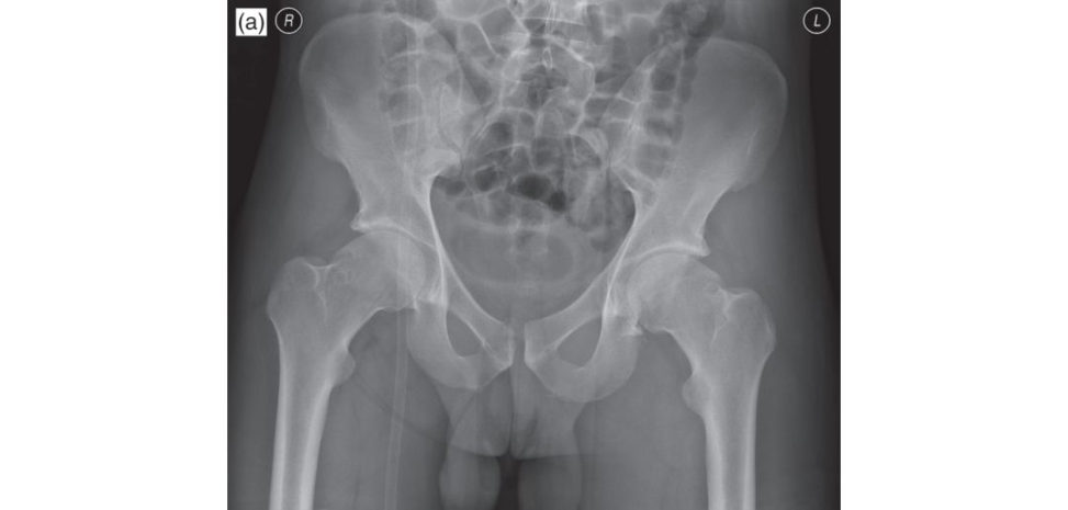
A 25-year-old professional motorbike racer came off his bike at more than 60 miles/hour speed. His only area of pain is his left hip. This is an X -ray of his pelvis. What does it show (Figure 14.1a)?
Anteroposterior pelvis radiograph of a skeletally mature adult marking out the iliopectineal and ilioischial lines (representing landmarks of the anterior and posterior columns) ... The medial aspect of the acetabulum is represented by the teardrop and the weight-bearing dome by the sourcil.
[Candidate prompt] Tell me about pelvic anatomy. What acetabular lines do you know and are any disrupted?
Practise out loud describing disrupted acetabular lines on pelvic radiographs until it all comes together. Big-volume trauma books or internet image searches are the best sources. The acetabular dome appears intact; Shent online is intact. My concern is a possible posterior wall injury. The right sacroiliac joint appears wider than the left. Both hip joints appear concentric.
What will you do next?
This is a high-energy injury and the patient should be assessed according to ATLS protocols so that life-threatening injuries are not missed. I will assess the range of movements in the left hip joint, distal neurovascular status and examine the left knee and left ankle. If, however the ATLS talk has already been done in the previous viva question then a candidate should default to ‘Assuming the injury is an isolated closed injury and ATLS protocols have been performed I will assess range of movements in the left hip joint ... etc.’
If exam tactics are really not your strongpoint, then if allthings fail at least you should avoid mentioning the ‘ATLS talk’.
Left hip movements are limited to a jog of movements by pain; the rest of the examination is unremarkable. What is the next step?
I will request a CT scan of the pelvis and both hips.
Would you order a CT scan in the middle of the night?
If the injury occurred during the day it should be fairly straighfoorward in most hospitals to obtain an urgent CT scan that day. If the injury presents in the middle of the night, say 2 a.m., it could wait until the following morning, as the scan does not need to be performed immediately. This question tests real-life decisions and the rationale (and evidence) behind your choice – what you will do in an actual situation with a real patient in front of you. This is the score 7 and 8 opportunity that if a viva gets stuck down on competency questions the candidate will never get to.
These are axial CT scans of both hips and SI joints. Describe the injury (Figures 14.1c and 14.1d).
The axial section of the left hip shows an intra-articular fragment, marginal impaction of the posterior wall, and loss of concentricity of the hip joint.
What is the definitive management of this injury?
The aims ofo per at ive management are to remove intra-articular fragments, reduce the marginal impaction, bone gr aft the bony defect if needed, then but iress plate fixation of the posterior wall.
When will you operate?
Surgery should be performed ideally within 5 days as per BOAST guidelines.
It is much better if a candidate avoids pure recitation of the BO AST.
What are BOAST guidelines?
British Orthopaedic Association Standards of Trauma guidelines.
Pelvic fractures
First line of management is control of haemorrhage – pelvic binder, blood transfusion, pelvic packing or embolization.
Look for genitourinary tract injury and open fractures – wounds in perineum, rectum or vagina.
Surgical treatment of these injuries as soon as possible.
Early CT scan of pelvis.
Transfer images to local referral unit within 24 hours.
Once haemodynamic and skeletal stabilizations are achieved, the patient should.
Figure 14.1a Anteroposterior (AP) radiograph of pelvis.
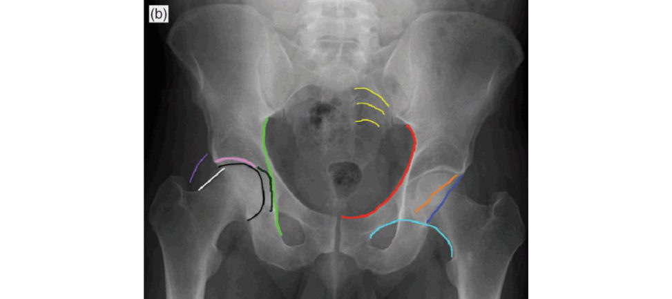
Figure 14.1b Iliopectineal liner ed); ilioischial line (light green); sacral arcuate lines (yellow); Shenton arc (light blueline of Klein (white); gluteal fat stripe (purple); acetabular roof (pink); medial acetabular wall (dark green); anterior acetabular wall (orange); posterior acetabular wall (dark blue); femoral headline (black).
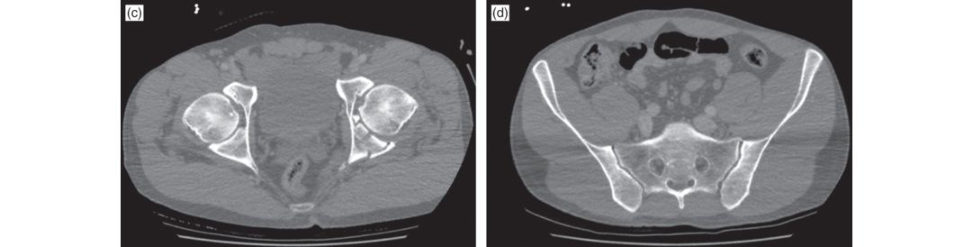
Figure 14.1c and 14.1d CT scan of pelvis and SI joints.
Acetabular fractures
CT scan within 24 hours and images should be transferred to the specialist unit.
Surgery if needed should be performed within 5 days, ideally.
What approach will you use? What are the significant risks and complications of the approach?
Posterior Kocher–Langenbeck approach. This allows access to the posterior wall and posterior column of the acetabulum.
Take me through this approach.
The skin incision begins 5 cm anterior to the PSIS, curves over the greater trochanter and runs parallel to the shaft of the femur for 15–20 cm. The deep dissection involves exposing the insertion of the piriformis tendon, the gemelli and the internal obturator muscle. The piriformis is divided through its tendon 1–2 cm from its femoral insertion after a stay suture has been passed through it. The tendons of obturator internus, superior and inferior gemelli muscles are tagged, divided 2–3 cm from their femoral insertion and then retracted. It is important to leave a cuff of tissue around the external rotators and avoid dissecting in to quadratus femoris in order to preserve the ascending branch of the medial femoral circumflex artery.
Would you prefer to position the patient in the lateral position or pr one?
This questionist esting higher -order thinking about what a surgeon would do in real life and what are his/her justifications for a particular decision. Higher rates of infection and revision surgery are reported in the prone group.
The main disadvantages of the lateral position are:
Difficulty applying manual traction.
Potential for sciatic nerve injury.
Difficulties achieving reduction duet o persistent posterior column.
Access through the greater sciatic notch for palpation or clamp placement is impaired.
The prone position is particularly indicated for transverse or T-type fractures. The leg can beheld flexed at the knee and extended at the hip to avoid traction on thes cia tic nerve, greatly reducing the chance of nerve injury.
The posterior column should be fixed first as it provides a stable surface to reduce the posterior wall fracture.
What are the significant risks and complications of the approach?
During surgery the short external rotators should be divided at least 1 cm from their insertions to protect this artery and avoid avascular necrosis of femoral head. It is important to make sure no screws are penetrating the joint using II or intraoperative radiographs. Other risks include infection, D VT, PE, loss of fixation heterotropic ossification and secondary osteoarthritis.
Can you think of any technical difficulties that you may encounter when fixing the fracture?
Traction is very important in allowing fracture reduction Traction unloads the joint allowing better joint visualization and assists direct manipulation of the fracture fragments. Bone graft may be needed to fill in any fracture gaps.
Does this injury have a good or a bad prognosis, historically?
Posterior wall fractures have in general poor prognosis due to the damage to.
What will be your postoperative rehabilitation protocol?
I will start hip range of movement exercises from day one and continue with non-w eight- bearing for 3 months.
Structured oral examination question 2#
A 23-year-old professional dancer is involved in a road traffic accident at 5 pm (motorbike rider vs. car). The patient is brought to casualty with GCS of 15, BP 110/70 mmHg, PR 90/min. The patient is complaining of pain around the right buft ock area. Fifteen minutes after arrival the patient’s BP dropped to 70 mmHg systolic. What will you do?
As per ATLS protocols I will perform primary and secondary survey making sure two large- bore cannulae are introduced and blood taken for FBC, U&E, cross-match 6 units of blood.
The patient blood pressure stabilized at 110/70 mmHg and 2 units of blood are being transfused. X-ray of pelvis was performed. Describe the injury (Figure 14.2a).
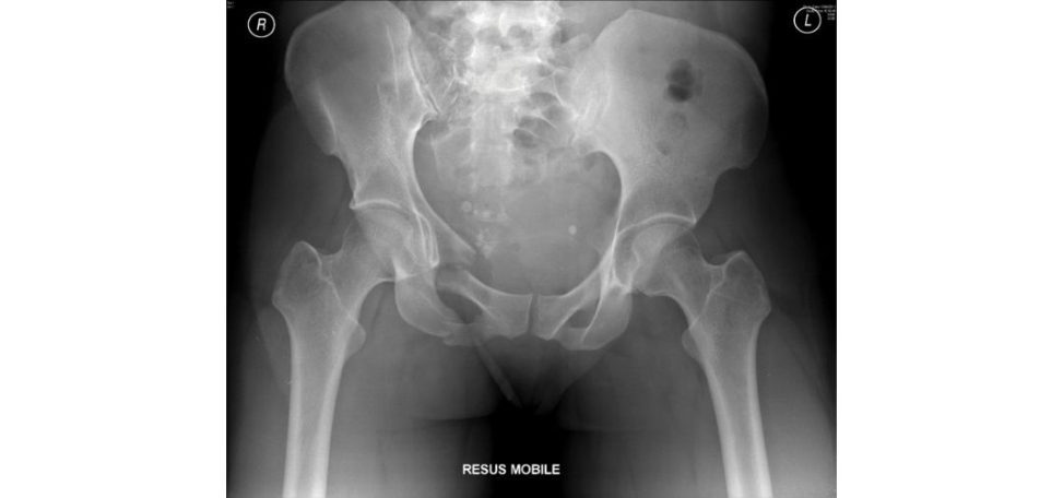
This is aver tical shear-type pelvic fracture involving the right hemipelvis with fractures.
Is there a spur sign?
The ‘spur’ sign represents the edge of intact ilium adjacent to the fracture, and is pathognomonic of a both-column fracture.
Is there any obturator ring disruption?
No.
Is there a fracture of the transverse process of L5?
I didn’t see one.
Why is this important?
A fracture of the transverse process of L5 in the presence of a pelvic.
You mentioned vertical shear, how can you classify pelvic injuries? CANDIDATE 1: [Silence ...] CANDIDATE 2: Judet and Letournel classification. I think you are mixing up acetabular and pelvic classification system names.
CANDIDATE 3: Pelvic fracture can be classified based on the stability of the pelvic ring.
Do you know a name?
Young and Burgess (Table 14.1).
How does this classification guide your management?
Fractures are divided into one of four categories based on the mechanism of injury, two of which are further subdivided according to the severity of injury.
What is the typical mechanism of injury for a lateral compression fracture?
[Long silence ... ]
What are the radiological landmarks/lines you assess for a pelvic fracture? Show them on the normal side.
For pelvic fractures I start looking at the pubic symphysis, pubic rami, iliac wing, sacroiliac joints, sacral alae, neural foraminae, sacral bodies, transverse processes of lower lumbar vertebrae, sacral spinous processes.
What is this view?
It demonstrates the anterior rim of the acetabulum and the posterior ilioischial column.
Can you identify the lines for me?
I am not sure.
Have a try.
Line one is ilioischial line, line 4 is the iliac crest.
How is this radiograph taken?
This is obtained on a supine patient with the injured side of pelvis rotated anteriorly at 45°.
That is the other Judet view, the obturator view.
The obturator oblique view is obtained on a supine patient with the injured side of pelvis rotated anteriorly at 45°. The X-ray beam is directed vertically toward the affected hip. The iliac oblique view is obtained on a supined patient with the unaffected side of the pelvis rotated anteriorly at 45°.
How will you manage the patient now?
I will also look for any open wounds around the perineum, groin, buft ocks vagina, rectum to rule out an open fracture.
The patient has altered sensation in the S1 nerveroot area of the right foot, but no motor deficit was noted. What do you do?
I will obtain CT scan of cervical spine, chest, abdomen and pelvis to assess for associated injuries and look specifically for any evidence of S1 nerveroot injury due to the pelvic fracture.
CT scan does not show any other visceral or vascular injuries. No urethral or perineal injuries were identified. What will be the definitive management and the timing?
Definitive management principles include reduction of the vertical shear, usually by skeletal traction, sacral fixation with sacroiliac screws, pubic ramus fixation with percutaneous ramus screw fixation or open reduction and plate fixation.
What are the specific risks involved?
Closed reduction of vertical shear may not be possible. The L5 nerveroot is at risk during sacroiliac screw insertion. Other risks include infection, D VT, PE, failure of fixation and persistent low back pain.
Figure 14.2a Anteroposterior (AP) radiograph of pelvis.
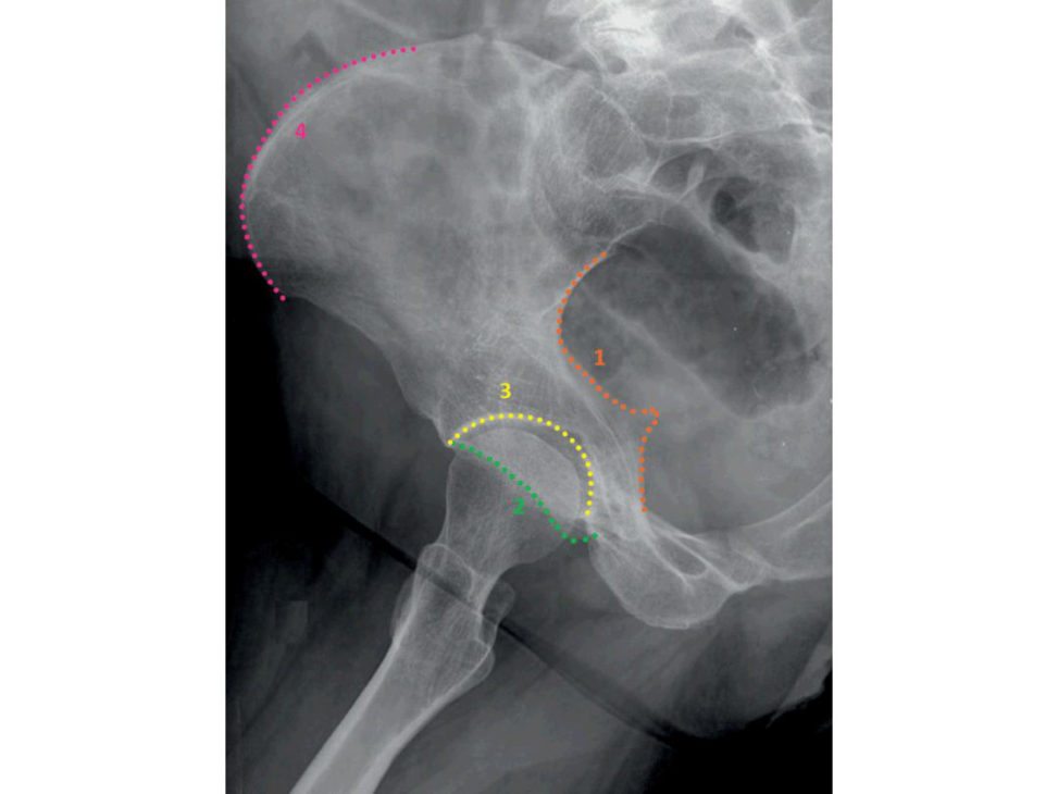
Figure 14.2b Radiograph of Iliac oblique view. 1, ilioischial line (posterior column); 2, anterior acetabular wall; 3, roof of acetabulum; 4, iliac crest.
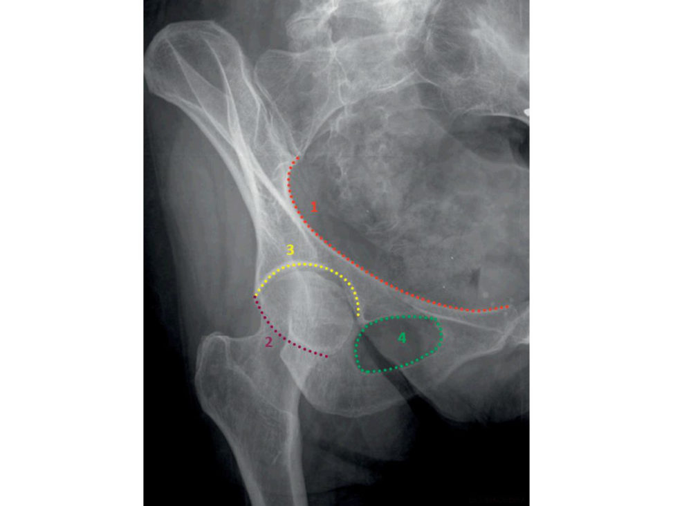
Figure 14.2c Obturator oblique view. 1, iliopectineal line; 2, posterior acetabular wall; 3, acetabular roof; 4, obturator foramen.
Table 14.1 Young and Burgess classification.
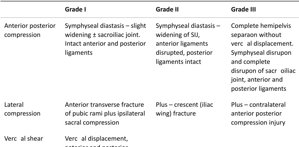
Vertical shear Vertical displacement, anterior and posterior through sacroiliac joint

Combination of other injury patterns: lateral compression/vertic al shear.
Structured oral examination question 3#
A 75-year-old gentleman who lives in a hostel, independently mobile, not on any medications sustained a fall while coming downstairs. He used to smoke 30 cigarettes a day and drinks ‘a lot’. This is an X-ray of his pelvis. Describe the injury (Figure 14.3a).
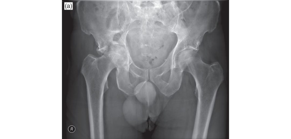
This is a right acetabular fracture with medialization of femoral head.
How will you manage this patient initially?
I will examine the pa tientas per ATLS protocols and make sure that there are no other injuries or any distal neurovascular deficits. I will apply distal femoral pin traction after making sure there is no femoral fracture.
Why do you want a CT scan? What is a CT scan going to tell you that a plain X-ray will not? Plain films were good enough 30 years ago before CT scanners were invented.
It also depicts soft -tissue complications, such as involvement of the sciatic nerve and the superior and inferior gluteal arteries.
What info will a CT provide that will change your management?
It will help you to identify the degree of fracture displacement more accurately.
CT scan axial views are shown in Figures 14.3b–14.3d. What type of fracture is it? How can you classify acetabular fractures?
Acetabular fractures can be classified by the Judet and Letournel classification. Posterior column.
There are also five associated patterns, which are a combination of the elementary patterns:
Posterior wall and posterior column.
Transverse and posterior wall.
T-shaped.
Anterior wall/column and posterior hemitransverse.
Bilateral column.
This fracture involves both anterior and posterior columns. There is an area of intact acetabular dome.
Hence, these fractures are often due to low-energy injuries, such as a fall from standing height.
What is your definitive management?
Non-operativ e management accepts some degree of malunion and if this becomes symptomatic a total hip replacement can be performed. Skeletal traction for 6 weeks followed by a further 6 weeks of non- weight-bearing but hip range of movements are started.
What are the advantages and disadvantages of just ORIF versus ORIF and THA?
However, apa tien t has to be non-weight- bearing for 3 months. Even if fixation does not fail, due to the increased risk of secondary osteoarthritis, a patient could still f ace a relatively big second operation Technically, total hip arthroplasty could be performed with relative ease as the fracture should have healed and will provide a stable base for the acetabular cup.
Figure 14.3a Anteroposterior (AP) radiograph of pelvis.
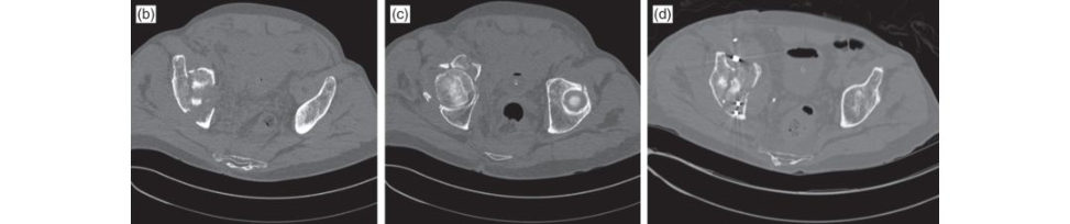
Figure 14.3b–14.3d CT scan axial views of pelvis.
Structured oral examination question 4#
A 48-year-old man known to have mental health issues jumped off a bridge from a height of 30 feet, landing on a concrete pavement. In A&E his injuries identified are all orthopaedic injuries. Lumbar spinal fractures at L2, L3 burst fractures, pelvic and hip injuries as shown in this X-ray (Figure 14.4a), fracture of left radius and ulna, closed intra-articular pilon fracture of left distal tibia. How do you manage this patient?
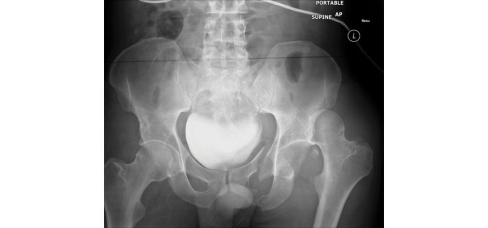
Closed reduction of left hip as soon as possible and distal femoral pin traction. Soft -tissues tatus of pilon fracture. Once haemodynamic stability is achieved, CT scan of neck, chest, abdomen and pelvis is performed to rule out other injuries and better identify the fracture patterns.
What are his injuries on X-ray?
Posterior dislocation of left hip with possible fracture, cannot say where the bony fragment has come from. Pubic rami fractures on left side.
Axial CT scans of pelvis are shown in Figures 14.4b and 14.4c. What do they show?
In conjunction with the pubic rami fractures, this is an unstable pelvic fracture.
What other reconstruction view is essential to look at?
The sagift al view of the sacrum will show whether there are any transverse sacral fractures.
What is the definitive management plan for this pelvic fracture and its timing?
The pelvic fracture fixation is withs pino pelvic stabilization with pedicle screws and a rod system connecting the fitih lumbar vertebra and posterior iliac spines followed by pubic ramus fracture fixation with plate and screws or percutaneous screw fixation.
Figure 14.4a Anteroposterior (AP) radiograph of pelvis.
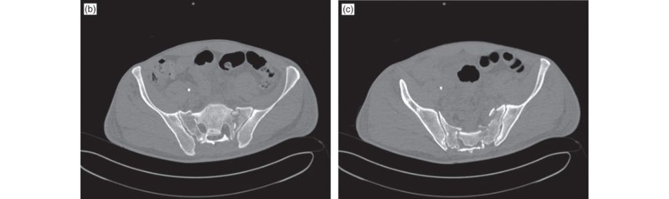
Figure 14.4b and 14.4c CT scan axial views of pelvis.
Structured oral examination question 5#
A 65-year-old lady front-seat passenger of a car involved in an RT Ais brought to A&E complaining of pain in her pelvic area and abdomen. GCS is 15, observations are stable. She is obese, suffers from hypertension, NIDDM, has had several laparatomies for diverticulitis, adhesions total hysterectomy. This is a reconstruction of a C T scan of her pelvis (Figure 14.5a). What is the fracture pattern?
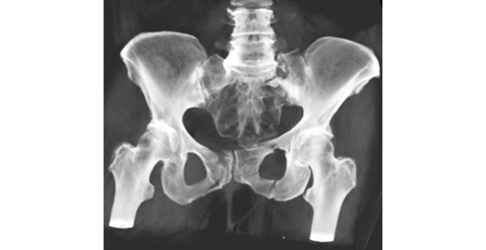
There is also vertical displacement along with fractures of left transverse process of fourth and fitih lumbar vertebrae. This is aver tic ally unstable fracture.
What are you looking for in examination of this patient?
As per ATLS protocols, I will perform primary and secondary surveys.
This is a CT scan axial view (Figure 14.5b). Describe the injury.
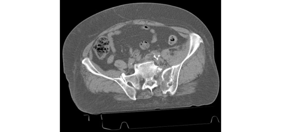
There is a fracture of the left half of the sacrum along the neural foraminae that is displaced.
Do you know of any classification for sacral fractures?
Type I – sacral ala fracture. Type II – fracture through neural foraminae. This fracture is Type II.
What is your management plan?
Left distal femoral pin traction. Transfer of images and patient information to specialist unit.
What are the options for managing the pelvic injury?
Anterior stabilization with plate and screws via Pfannesteil approach.
What peri operative difficulties do you anticipate?
If posterior transiliac rods fixation is planned, then positioning the patient may be difficult.
Intraoperative fluoroscopic images will be suboptimal duet o obesity.
Poor bone quality with poor bone purchase of screws.
Due to previous abdominal procedures, exposing pubic ramus and symphysis.
Figure 14.5a Anteroposterior (AP) radiograph of pelvis.
Figure 14.5b CT axial view of pelvis.
Structured oral examination question 6#
A 16-year-old male pedestrian was hit by a car at about 40 miles/hour speed. GCS at scene was 5–6. Hence, he was intubated at scene. Systolic blood pressure is around 90 mmHg, PR 100/min and peripheral pulses are well felt. Trauma series show no chest or neck injury, but pelvic X-ray has been taken. Describe the injury (Figure 14.6a).
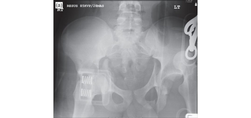
There is a posterior dislocation of the left hip with associated acetabular fracture and anterior dislocation of right hip with associated acetabular fracture.
CT scan of head, neck, chest, abdomen, pelvis was done. It showed cerebral oedema, fluid in the abdomen and the injury to both hips as seen in Figures 14.6b–14.6d. How will you manage the orthopaedic injuries?
Both hip dislocations require urgent reduction and regular check of distal vascular status.
What are you worried about?
The right femoral head is probably very close to the external iliac artery.
After closed reduction of both hips what will you do?
I will perform distal femoral pin traction for both lower limbs, organize a CT angiogram to confirm the patency of the external iliac artery even if there are good pulsations distally.
What will be your definitive management?
When the patient is safe for transfer, patient will undergo open reduction and internal fixation of both acetabular fractures either in the same siting or asas taged procedure.
Figure 14.6a Anteroposterior (AP) radiograph of pelvis.
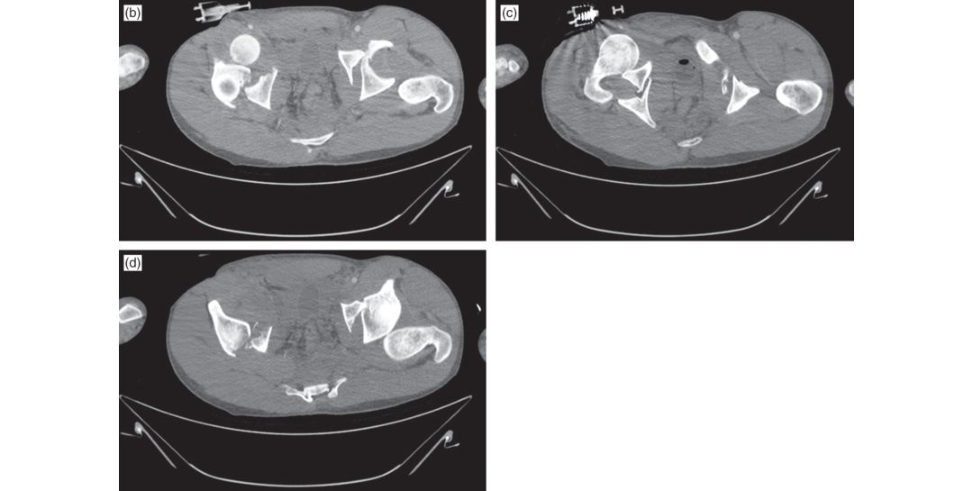
Figure 14.6b–14.6d CT scan axial views of pelvis.
Structured oral examination question 7#
A 29-year-old male cyclist has been admift ed to casualty after being hit by a car. A trauma call has been putout. He has had pelvic radiographs taken by the A&E team involved with his initial c are (Figure 14.7a). What do they show?
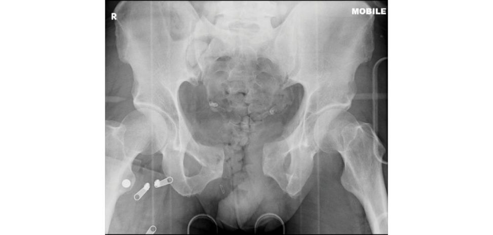
This is an AP pelvic radiograph which shows an open-book pelvis fracture with diastasis of the pubic symphysis. This is a significant injury usually sustained through high-energy trauma and is often associated with other severe injuries.
What are the ATLS principles?
The primary survey involves: (1) Assessment of airway with cervical spine control. (2) Breathing and ventilation. (3) Circulation with haemorrhage control. (4) Disability/neurologic assessment. (5) Exposure of the patient. The secondary survey is a ‘head to toe’ evaluation of the patient including full history, examination and reassessment of all vital signs.
Who would you want to be in the trauma team? Who should lead the team?
The t eam leader should ideally be an Accident and Emergency Consultant. The Airway doctor should be someone competent in difficult airway management, usually an anaesthetist These doctors should each have a nurse assisting them.
You mentioned a pelvic binder earlier , how would you apply this?
The pelvic binder is positioned a t the level of the greater trochanters and is tightened at this level.
What could you do if one is not available?
The aim of this or the binder is to splint the bony pelvis and reduce haemorrhage from venous disruption.
The patient is found to be profoundly hypotensive despite fluid resuscitation. It is f elt the patient is bleeding into their pelvis and it is suggested that the major haemorrhage protocol is activated What do you know about this protocol?
The major haemorrhage protocol varies slightly from hospital to hospital but is activated when apa tien t is identified who would benefit from a large-volume blood transfusion. These should be transfused in a 1:1:1 ratio to prevent coagulopathy following large-volume transfusion.
Is your hospital 1:1:1?
I think my own hospital is 2:1:1 and I think most hospitals, unless an MT Care 4:4:1.
How does tranexamic acid work?
It acts as an anti fibrinolytic by reversibly binding to lysine receptor sites on plasminogen.
Figure 14.7a Anteroposterior (AP) pelvic radiograph demonstrating open-book pelvis.
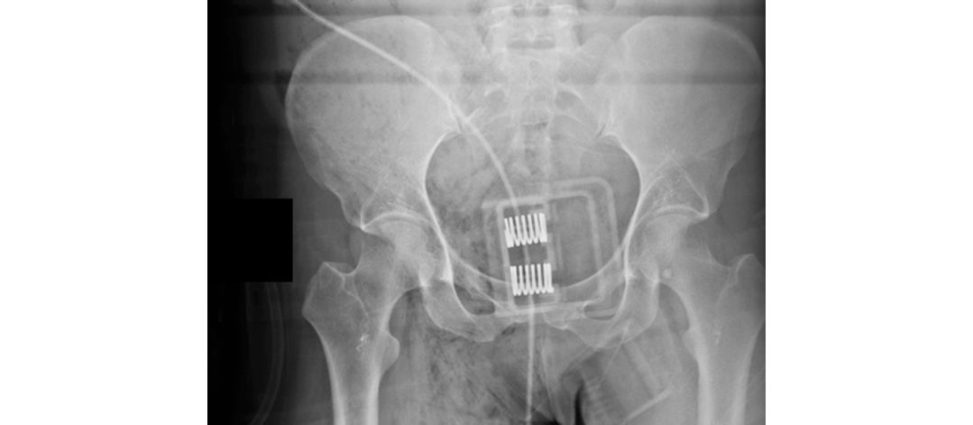
Figure 14.7b Radiograph of pelvic binder in situ. Candidates should be able to recognize binder placement on a pelvic radiograph and if incorrect position.
The CRASH trial showed that it reduced mortality.1
How much tranexamic acid?
1 g IV loading dose within 3 hours of injury followed by a second dose 1 g over 8 hours.
Whatabout crystalloids and vasopressin?
I would avoid using them.
Why?
Because they interfere with the resuscitation process.
How?
Large amounts of early high-dose crystalloids can cause an imbalance of coagulation haemostasis. Giving blood early is a better alternative and a move away from the traditional A TLS teaching of 2 lof crystalloids stat.
What is permissive hypotension?
This refers to managing trauma patients by restricting thea mount of resuscitation fluid and maintaining.
Do you know any common complications from massive transfusion?
Hypothermia, thrombocytopaenia from dilution metabolic alkalosis and hypocalcaemia due to.
Despite this, the patient continues to remain hypotensive. There are no other obvious injuries apart from the pelvis injury. How would you manage this patient now?
The patient could either betaken to theatre and pelvic packing performed in order to tamponade the bleeding. The choice between the two would depend on resource availability and surgical experience.
Selective or non-selectiv e embolization?
I would definitely prefer selective embolization.
Why?
Non-selectiv e embolization may lead to an increased risk of wound-healing complications, increased risks of hip ON, fracture non-union peripelvic soft -tissue necrosis and infection You shotgun the internal iliac artery and are causing a wide ischaemic insult to the surrounding soft tissues with an increased risk of severe sepsis occurring.
What about genitourinary injury?
Widening of the symphysis pubis and sacroiliac joint may predict bladder injury while fractures of the inferior and superior pubic rami are more commonly associated with urethral injuries. Clinical examination may reveal bleeding from the urethral meatus.
Structured oral examination question 8#
You are called totheR esus department to see this 23-year-old who has been hit by a bus while drinking on a night out. His pelvic radiograph is shown here (Figure 14.8a). What does it show?
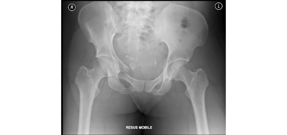
This AP pelvis radiograph shows a lateral compression (LC) type pelvis fracture, with fractures to the pubic rami and ipsilateral posterior ilium.
What are your priorities for this patient?
I would want a warm, oxygenated and well-perfused patient. This is the ATLS talk but in a subtler, less obvious manner.
What else?
I would activate the trauma team.
Who is in the trauma team?
The trauma team is a multidisciplinary team consisting of doctors and nurses from different specialties (see previous question).
It should be consultant-led because this saves lives. What is a sterile handover?
This is where the ambulance crew hand over the patient to the trauma team.
What else?
The ambulance crew provide a detailed handover of the patient in a quiet area of casualty and the trauma team refrain from treating the patient unless there is an impending airway problem or visible catastrophic haemorrhage.
What do we mean by ATMIST?
This stands for: Age. Time of incident. Mechanism of injury. Injuries top to toe. Vital signs.
Mode of transport.
Typically, what other injuries are associated with this type of fracture?
This differs to open-book AP compression type fractures which are typically associated with urethral and bladder injuries and pelvic vascular injuries.
You arrive in the emergency department and his observations are: pulse 120, BP 95/55 with a capillary refill time of 3 seconds and feels cool peripherally. He is confused and agitated. What are your thoughts?
Shock is inadequate perfusion of tissues and is an emergency. His confusion and agitation may be due to hypoperfusion of the brain secondary to his hypovolaemic state, or it may be due to an associated head injury. My priorities are the management of immediate life-threatening injuries.
How can shock be classified?
Classes 3 and 4 are life-threatening and require blood product transfusion. Other causes of shock include neurogenic shock, septic shock and haemodynamic shock from causes such as cardiac tamponade and tension pneumothorax.
An AB Gis carried out and his lactate is found to be 4.1 mmol/l. What is the significance of this?
Lactate levels below 2.5 mmol/l are generally accepted to show adequate resuscitation. Anything above 4 mmol/l would be an indication for damage control orthopaedics (DCO) while between 2.5 and 4 mmol/l the patient needs to have a period of resuscitation before considering surgery. Below 2.5 mmol/l is usually good for surgery, but this is based on a trend.
In particular , no single physiological parameter or blood marker can as yet be used to guide intervention, but the accepted level of 2.5 mmol/l for lactate is likely too conservative and is being superseded by a more comprehensive and patien t-centred approach, focusing on physiological improvement and reversal of acidosis reflected by a lactate < 4.0 mmol/l, pH ≥ 7.25, or BE above 5.5 mmol/l.
On the same ABG the patient’s O2 saturation was 91%. What is the significance of this?
A normal value in an individual with no underlying lung disease is above 96%. At levels below 96%, due to the sigmoid shape of the oxygen dissociation curve, the pO2 drops quickly for a decreasing oxygen saturation reading.
You have given him 2 units of blood, his pulse is 120, BP 95/60, O2 saturations 94% on high- flow oxygen. What now?
I would want to identify any injuries directly related to the pelvic fracture.
Why are you applying a pelvic binder? It isn’t an open-book pelvis. Will it help you with resuscitation of the patient?
Pelvic binders control bleeding by compressing and stabilizing fractures, not by significantly reducing pelvic volume.
What about worsening an injury when a pelvic binder is applied?
As a fracture pattern is often unknown before pelvic binder application, it is possible to exacerbate certain injury patterns if excessive force is applied.
What are the characteristics of the ideal binder?
The ideal pelvic binder: Should belight; Allows access to the abdomen for laparotomy.
May need to stay on for 24 hours or more and thus should be of a.
Should not limit access to the perineum and anus for examination.
Must fit various sizes of patients including children).
Should be washable or cheap enough to be disposable.
How long would you leave a pelvic binder on for?
Up to 24 hours.
That is probably too long. One would beworried about pressure sores and skin abrasions. What is the function of the pelvic binder?
[Silence ... ] To stop bleeding.
To allow the unhindered formation of clot which wills top the bleeding. If you disturb the clot re-bleeding will occur. In our hospital we aim to have released the pelvic binder by 8 hours. We would make sure the patient is normothermic, haemodynamically stable and any metabolic acidosis corrected. All imaging should have been completed and the fracture should have been fully characterized, and we have a definitive plan of action in place for management if the patient becomes unstable once more.
The examiner wouldn’t necessarily volunteer any of this info unless it was a mock examination.
How do you determine if there is a coagulation issue?
I would check full blood count, U&Es and coagulation screen.
Anything else?
Ensure patient warming, avoid acidosis and perform an INR.
Have you heard of thromboelastography (TEG) and rotational thr omboelastometry (ROTEM)?
No. The candidate isn’t scoring well in the viva (possibly 4 or 5). TEG allows real-time assessment of cloting and may be a valuable adjunct in acute resuscitation and guide transfusion of blood products different from a standard 1:1:1 ratio.
What next?
I would consider obtaining a CT angiogram to investigate for ongoing sources of bleeding.
What type of CT angiogram?
A triple-phase CT contrast angiogram is my modality of choice. The three phases are arterial, portal venous and delayed phase.
You don’t have access to a CT angiogram as it wouldn’t be ready for at least 2 hours as the vascular list has overrun.
As the patient is haemodynamically unstable he will need to go to theatre for more definitive pelvic stabilization and for pelvic packing.
What type of pelvic packing?
Extraperitoneal.
What else?
The vast majority of pelvic bleeding originates from the presacral venous plexus and fracture.
How do you perform pelvic packing?
I have never seen it performed, but the packing is into the preperitoneal pelvic packing.
What incision would you use?
Aver tical incision is preferred to a Pfannenstiel incision.
Why?
For PP Paver tical midline incision is made from the pubic symphysis extending up 6–8 cm. The laparotomy incision may extend from the xiphoid to just below the umbilicus while the PPP incision is approximately 6 cm away in the suprapubic area. Packs need to be removed after 48 hours.
Figure 14.8a Anteroposterior (AP) radiograph pelvis demonstrating lateral compression injury.
Notes
1. A bland, throw-away statement that doesn’t score a candidate any points.
2. Much better and more strategic than 1.