Chapter 17 Hand and upper limb
Structured oral examination question 1#
Distal radius fracture and malunion
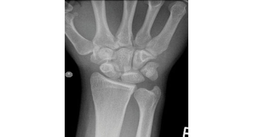
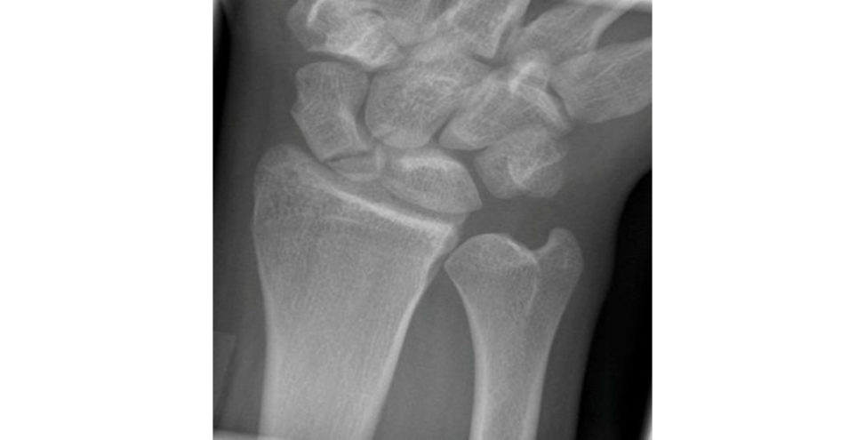
Please describe the radiographic findings (Figure 17.1a and 17.1b).
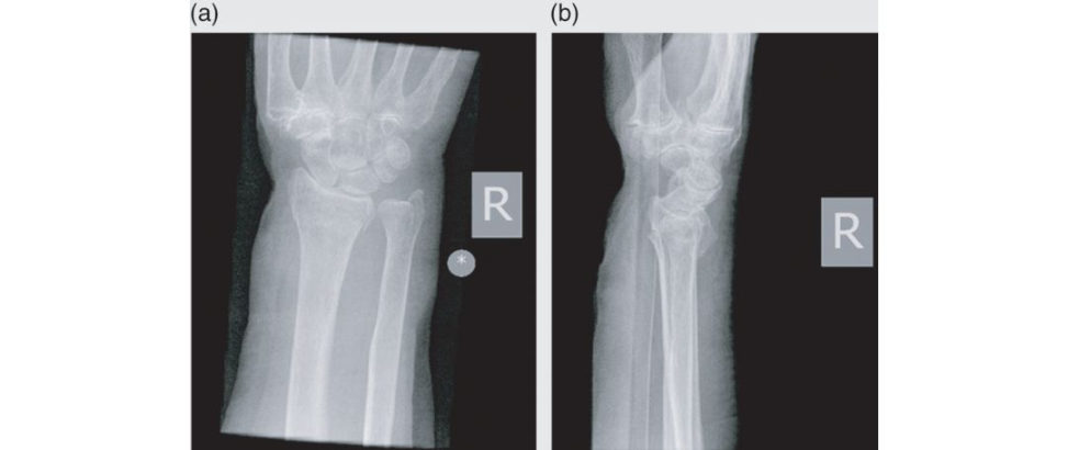
These are posteroanterior (PA) and lateral radiographs of the wrist that show an extra- articular distal radius fracture. The radiographs also showed thumb carpometacarpal arthritis.
These are the radiographs of an 83-year-old lady. What are the acceptable parameters for conservative management of distal radius fracture post manipulation in A&E?
I would look at the overall alignment of the distal radius including less than 10° dorsal angulation of the distal radius articular surface; if this was an intra-articular fracture then less than 2 mm gap or step off, radial length within 2 mm of the ulna length, radial inclination of 21°, and no secondary carpal malalignment.
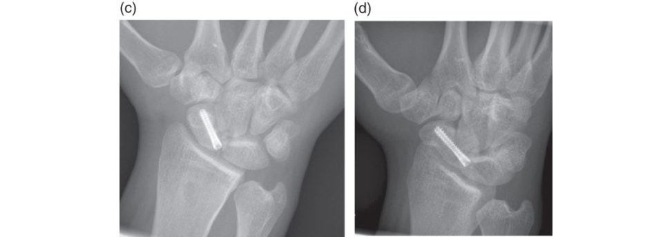
The on-call registrar has kindly reduced this fracture in A&E under haematoma block. The position in belo w-elbow plaster post closed reduction a t 1 and 2 weeks follow-up in the fracture clinic was acceptable. Please comment on these radiographs taken at 6-week follow-up in the fracture clinic following conservative management of the distal radius fracture (Figure 17.1c and 17.1d).
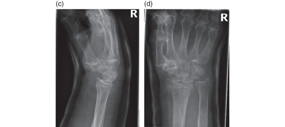
These are posteroanterior (PA) and lateral radiographs of the wrist in a plaster that show extra-articular distal radius fracture.
What would you do now?
I would inform the patient that the fracture has slipped and healed in a less-than-ideal position. I would arrange urgent physiotherapy in order to maximize her rehabilitation. I would review her progress in clinic in 6 weeks with repeat radiographs.
The patient asks why the fracture wasn’t fixed surgically.
I would mention to the patient the complications that could occur with volar locking plate fixation including infection, painful scar, tendon rupture/irritation, injury to neurovascular structures such as the median nerve and radial artery, screw cutout and hardware failure, carpal tunnel syndrome, secondary fracture displacement and chronic regional pain syndrome (CRPS).
Don’t forget to mention CRPS as a possible complication from any type of hand surgery undertaken.
That’s assuming you would have treated this fracture with a volar locking plate and not K- wire fixation, but w e’ll not go there.
The management of displaced distal radius fractures is controversial. A recent meta-analysis showed no significant difference between patients who underwent internal fixation and conservative management for displaced distal radius fracture sUK DRAFFT).
The patient comes back in 8 weeks and now complains of difficulty performing her daily tasks due to the wrist deformity. What would you offer her at this stage?
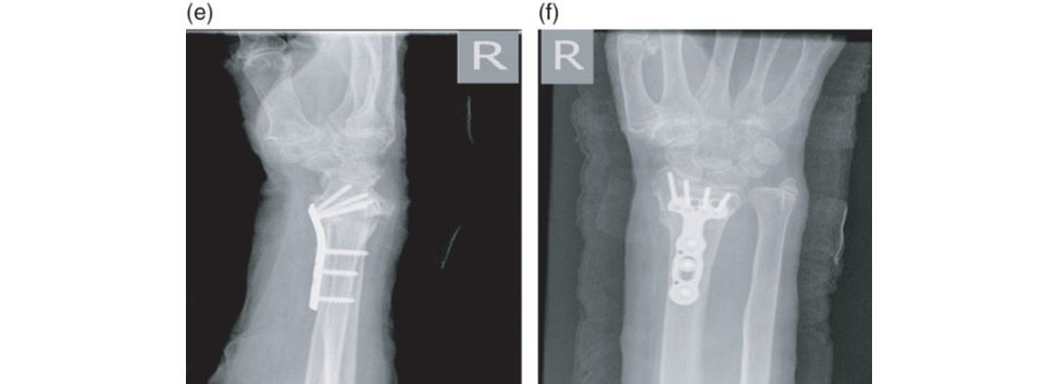
If the patient had ulnar- sided wrist pain and due to shortening from the distal radius malunion, I would offer her a correction osteotomy of the distal radius using a volar locking plate through an extended flexor carpi radialis (FCR) approach. I would immobilize the wrist in a short arm plaster for 2 weeks for wound healing. At 2 weeks I would change it to a future-type wrist splint to allow mobilization of the wrist once the plaster is removed, 4 times a day, and take a check X-ray at 6 weeks to confirm bony union (Figure 17.1e and 17.1f). Another scenario: apa tien t with distal radius fracture treated non-operativ ely came back at 8 weeks with persistent pain, swelling and erythema.
How would you approach this patient?
I would like to take a thorough history and examination of the wrist.
What is chronic regional pain syndrome?
Chronic regional pain syndrome (CR PSis contingent on the presence of regional pain combined with autonomic dysfunction, a trophy and functional impairment affecting musculoskeletal, neural and vascular structures.
Type 1 – initiated by trauma with no identifiable peripheral nerve injury.
Type 2 – associated with identifiable peripheral nerve injury after trauma (causalgia).
How do you confirm the diagnosis?
CR PSis principally a clinical diagnosis based on the patient history and physical examination. There are no specific tests to confirm CRPS. The clinical presentations of CRPS can be divided into acute and chronic.
How are you going to treat the patient?
I will make an urgent referral to the pain team. Other treatments that may be considered include sympathetic nerve block and medical treatment (local anaesthesia, anft-epilep tics and anft-depr essants).
Figure 17.1a and 17.1b Posteroanterior (PA) and lateral radiographs of extra-articular distal radius fracture.
Figure 17.1c and 17.1d Posteroanterior (PA) and lateral radiographs of malunited distal radius fracture.
Figure 17.1e and 17.1f Posteroanterior (PA) and lateral radiographs of the wrist showing correction osteotomy with autologous bone graft.
References#
Ng CY, McQueen M. What are the radiological predictors of functional out come following fractures of the distal radius? J Bone Joint Surg Br. 2011;93(2):145–150.
Costa ML, Achten J, Plant C, et al. UK DRAFFT: a randomised controlled trial of percutaneous fixation with
Kirschner wires versus volar locking-plate fixation in the treatment of adult patients with a dorsally displaced fracture of the distal radius. Health Technol Assess. 2015;19(17).
Leung F, Kwan K, Fang C. Distal radius fractures: current concepts and management. Focus on series, Bone
Joint. 2013.
Structured oral examination question 2#
Extensor pollicis longus (EPL) tendon rupture
What does the photograph (Figure 17.2a) show?
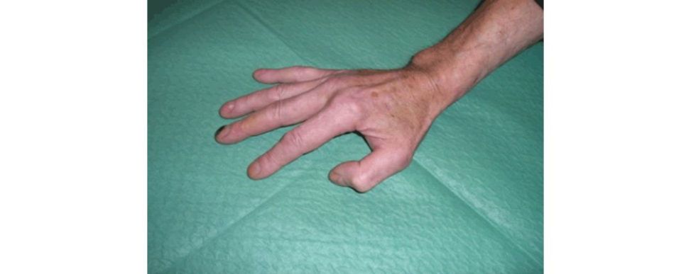
This is a clinical photograph of the right hand with the thumb in an abnormally flexed posture at the interphalangeal (IP) joint.
The patient has recently come out of plaster for a distal radius fracture. What is the likely pathology?
This is usually caused by rupture of the extensor pollicis longus (EPL) tendon at the level of the Lister’s tubercle in the third dorsal extensor compartment at around 6–8 weeks. EPL rupture usually occurs after an undisplaced or minimally displaced distal radius fracture. It occurs in about 1% of cases secondary to either atirition or ischaemia. It can bethought of as a type of compartment syndrome occurring in the tendon tunnel from fracture bleeding leading to interruption of tendon blood supply, nutrition and eventual rupture.
How would you test fo rEPL rupture?
The patient would be unable to extend the IP joint of the thumb.
I would not rely on testing for thumb IP extension as the EPB tendon inserts into the extensor apparatus of the thumb at varying levels and may be able to extend the IP joint of the thumb. Any other tests you would perform?
I would ask the patient to lift their thumb off a t able, which is not possible without an intact EPL tendon.
Can you describe the extensor tendon compartments at the wrist?
Abductor pollicis longus (APL) – attaches to thumb metacarpal. Second dorsal compartment (intersection syndrome): Extensor carpi radialis brevis (ECRB) – attaches to middle finger metacarpal.
Extensor carpi radialis longus (ECRL) – attaches to the index finger metacarpal.
Third dorsal compartment (passes around Lister’s tubercle):
Extensor pollicis longus (EPL) – passes around Lister’s tubercle of radius and inserts on distal phalanx of thumb.
Fourth dorsal compartment (contains PIN on the floor of this compartment):
Extensor indicis proprius (EIP) – lies deep to EDC tendon.
Extensor digitorum communis (EDC) – no direct attachment to proximal phalanx, attaches to the extensor expansions.
Fitih dorsal compartment (approach to DRUJ through floor of the fitih compartment):
Extensor digift minimi (EDM) – attaches to extensor expansion of litile finger.
Sixth dorsal compartment:
Extensor carpi ulnaris (ECU) – attaches to base of litile finger metacarpal.
How do you manage EPL rupture as in this case?
I would like to perform a functional assessment of the affected hand to determine the functional loss and deformity from the EPL rupture. At this stage I would refer the patient to a hand surgeon for surgical reconstruction. This would need reconstruction with tendon transfer, normally extensor indicis proprius to restore extension movement of the thumb.
What is the role of ultrasound scan in EPL rupture?
Sonography is to confirm the diagnosis of an EPL tendon rupture.
You are the hand surgeon, how many incisions would you use? Can you show it on my hand?
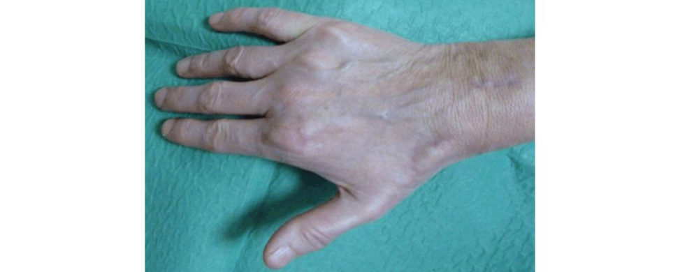
The extensor indic isis sutured to the remnant of the EPL tendon distally using the Pulvertaft w eave technique (Figure 17.2b).
How do you test for extensor indicis preoperatively?
Point the index finger with the middle to litile fingers fully flexed (this prevents EDC acting).
What are the basic principles of tendon transfer?
Principles when deciding on tendon transfers are:
1. The donor tendon must match the muscle strength.
2. The force of donor tendon should be proportional to muscle cross-sectional area.
3. Work capacity = force × amplitude.
4. Amplitude should be proportional to length of the muscle.
5. Motor strength will decrease one grade after transfer.
6. Appropriate tensioning of the donor tendon.
7. Appropriate excursion of donor tendon (can adjust with pulley or tenodesis effect).
The other requirements include patient compliance, no joint contractures,
The other variant of this question is if an extensor tendon rupture has occurred following volar locking plate fixation Typically, this would be a comminuted displaced intra-articular fracture treated with volar locking plate.
How will you minimize the risk of extensor tendon rupture following volar locking plate fixation?
The surgeon should choose the appropriate length of screw to avoid penetration of the far cortex causing tendon irritation and rupture.
Are there any other causes of EPL rupture?
EPL rupture can occur secondary to rheumatoid arthritis, bony spur developing after a.
Figure 17.2a Clinical picture of a hand.
Figure 17.2b Clinical picture of a hand showing restoration of thumb extension following reconstruction of EPL. Note the three incisions used.
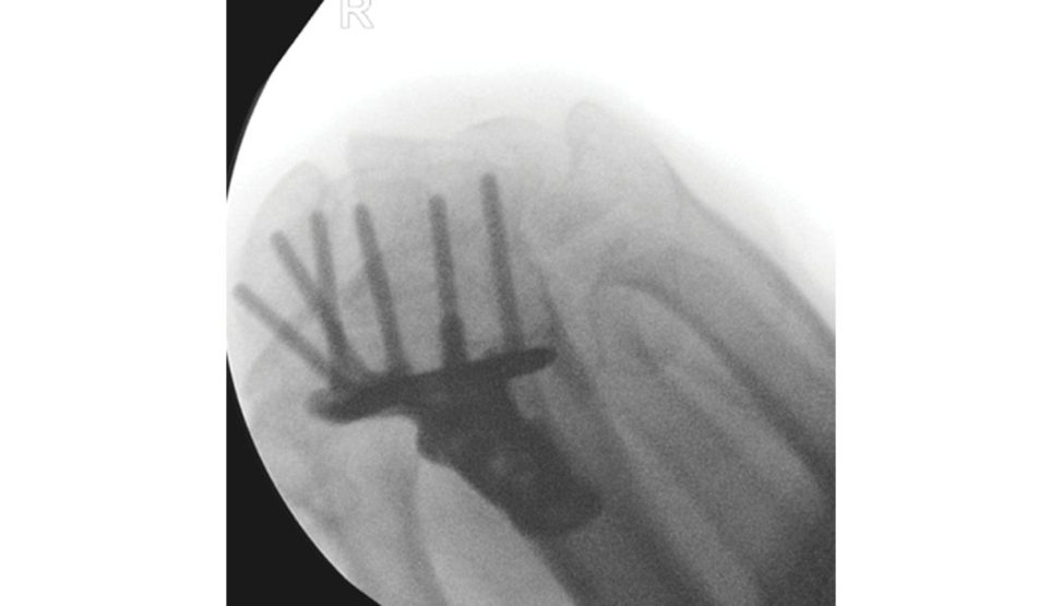
Figure 17.2c Skyline view distal radius taken during a volar locking plating with wrist maximally flexed. Note: the most radial peg has probably just penetrated through the cortex.
Structured oral examination question 3#
Transscaphoid perilunate fracture–dislocation
Please describe this X-ray (Figure 17.3a).
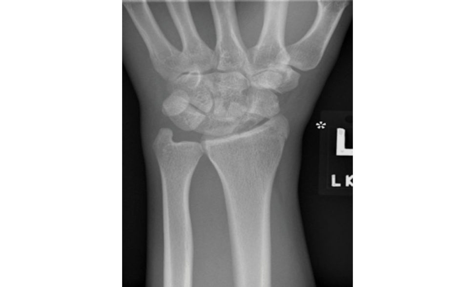
This is a posteroanterior view of the left hand showing transscaphoid perilunate fracture– dislocation.
Here it is (Figure 17.3b).
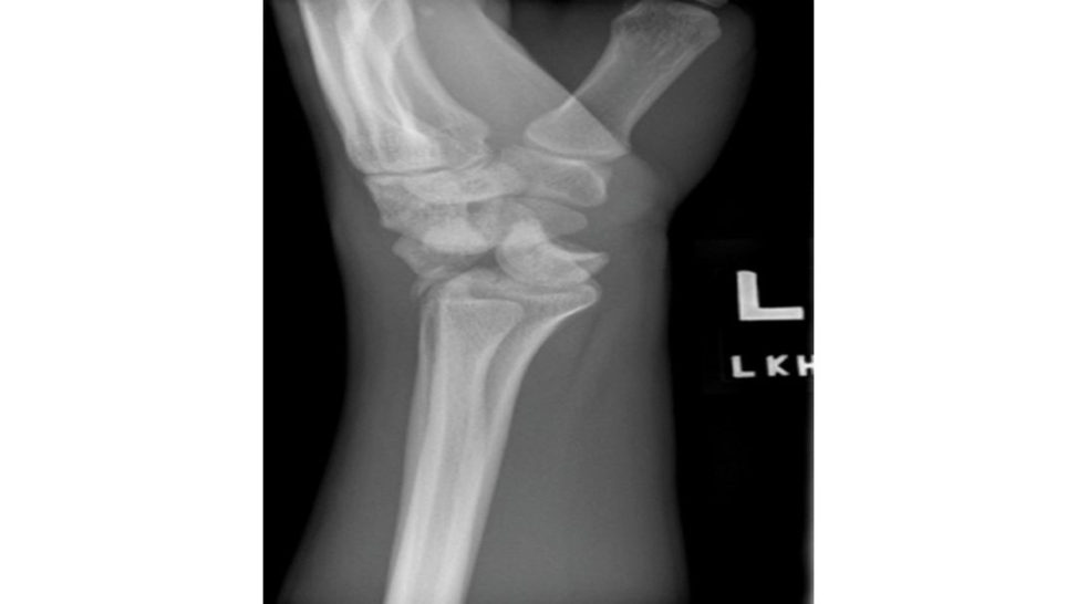
This lateral radiograph shows a perilunate dislocation with dorsal dislocation of the carpus. This is a greater arc injury and stage III on the Mayfield classification. Mayfield classification has divided this injury into four sequential stages. Stage I – scapholunate dissociation. Stage II – lunocapitate disruption capitate dislocates). Stage III – lunotriquetral disruption ( ‘perilunate’). Stage IV – lunate dislocation from lunate fossa (usually volar). Lesser arc injuries refer to purely ligamentous wrist injuries.
What is the difference between perilunate dislocation and a trans scaphoid perilunate dislocation?
A transscaphoid perilunate dislocation is the same injury but with an associated scaphoid fracture.
How would you like to proceed with this patient?
Assuming that ATLS has been performed in the initial management and life-threatening injuries have been excluded, I would like to proceed and take a thorough history including hand dominance, occupation and mechanism of injury. I would examine the hand, looking for deformity, swelling, open wound, perfusion status and check for median nerve symptoms. This would need to be reduced as an emergency in theatre with an image intensifier as prolonged dislocation increases swelling to the area and is associated with a higher rate of nerve injuries.
You have been successful in performing the closed reduction. How would you proceed now?
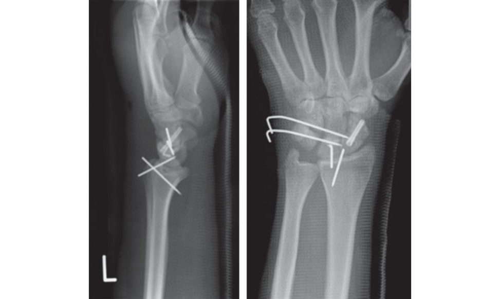
I would request an MRI scan to confirm the extent of injury as part of preoperative planning. The patient would need to have open reduction and internal fixation of the scaphoid and reconstruction of the sc apholunate and lunotriquetral ligaments through a volar or dorsal wrist approach. The dorsal approach is favoured by many as it provides access to the midcarpal joints so that intrinsic ligaments can be repaired. K-wire fixation is s till routinely used Scaphoid fractures are generally fixed with a headless compression screw. Stabilization of the lunate and reconstruction of the sc apholunate ligament are frequently performed using suture anchors (Figure 17.3c).
What is the expected outcome following this injury?
Patients should be counselled following this injury that there is a high risk of long-term problems of pain, stiffness and pos t-traumatic arthritis (36%). Carpal instability is also linked with this injury.
What is the normal scapholunate angle?
The average scapholunate angle is 45° (abnormal if < 30° or > 60°).
What is the ‘spilled tea-cup sign’ (Figure 17.3d)?
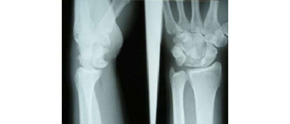
The spilled tea-cup sign describes abnormal volar displacement and tilt of a dislocated lunate on a lateral radiograph of the wrist. It is an important sign to help differentiate lunate dislocation from perilunate dislocation.
Figure 17.3a Posteroanterior (PA) wrist view demonstrating trans scaphoid perilunate fracture– dislocation.
Figure 17.3b Lateral wrist view demonstrating trans scaphoid perilunate fracture–dislocation.
Figure 17.3c Postoperative radiographs showing ORIF of the scaphoid and repair of capitolunate and lunotriquetral ligaments (note the bone anchor in the lunate) and temporary K-wire stabilization of the carpus, all done through a dorsal approach.
Figure 17.3d PA and lateral radiographs demonstrating a lunate dislocation (spilled t ea-cup sign).
Structured oral examination question 4#
Scaphoid fracture
You saw a 44-year-old patient in the fracture clinic with the above radiographs 2 days following an injury. Please comment on these radiographs (Figure 17.4a and 17.4b) and how you would manage this patient.
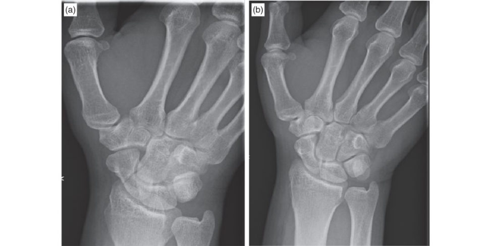
The radiographs taken show a scaphoid waist fracture. I would take a thorough history and examination of the hand and wrist. I would like to know the handedness, occupation, hobbies and previous similar injury in the history. If these confirmed the fracture was undisplaced, I would manage this non-operativ el yin a short arm cast without thumb immobilization for 6 weeks initially . The incidence of non-union at 6 months follow-up is independent of type of cast used. The healing rate for conservative treatment is 80–90% for undisplaced waist of scaphoid fracture.
What is the blood supply to the scaphoid?
Between 70% and 80% of the intraosseous vascularity and the blood supply to the entire proximal pole enters this way with no blood vessels penetrating the cortex of the proximal pole separately, explaining the higher rate of avascular necrosis with proximal pole fractures [2,3].
What are the indications for internal fixation of the scaphoid?
The indications for internal fixation of scaphoid fracture include displacement of more than 1 mm, intrascaphoid angle > 20° (humpback deformity), delayed union, proximal pole fracture or scaphoid fracture associated with perilunate dislocation. The indications for internal fixation of the scaphoid remain debatable.
These are the patient scaphoid radiographs at 4 months follow-up in the fracture clinic (Figure 17.4c and 17.4d). He remains symptomatic. What would you do?
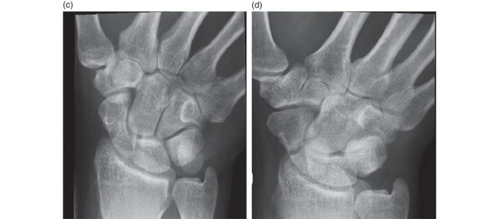
I would like to request a computed tomography (CT scan) to confirm the diagnosis of non-union, which forms part of preoperative planning. I would offer the patient bone gratiing and surgical fixation with a headless compression screw through either a volar approach if the CT scan confirms a humpback deformity, or the dorsal approach if there was no carpal malalignment.
The volar approach to the scaphoid utilizes the FC Ras an anatomical landmark with incision extended 2 cm proximal to the scaphoid tubercle with the distal incision directed towards the base of the thumb. The FCR sheath is then opened as distally as possible and retracted ulnarly to protect the radial artery and retracted radially to expose the capsule. The capsule and intracapsular ligaments are carefully divided and reflected off the scaphoid.
The dorsal approach is the preferred technique for proximal pole scaphoid fractures. The structure at risk is the superficial radial nerve branch. The extensor retinaculum is identified and incised, taking care to protect the EPL.
The patient has heard about percutaneous fixation techniques. What will you say to the patient?
Haddad reported the results of 15 patients with minimally displaced or non-displaced scaphoid fractures treated percutaneously using a volar approach, traction and a cannulated screw [7]. The early rigid fixation of scaphoid fractures allows early mobilization, patients were allowed movement soon after operation. The range of movement after union was equal to that of the contralateral limb and grip strength was 98% of the contralateral side at 3 months.
What are the complications of percutaneous fixation techniques?
The complications of percutaneous fixation techniques can be divided according to the approach being used, either the volar or dorsal approach. Possible complications for both approaches include guide wire breakage, prominence of the screw (through the subchondral bone), bleeding, infection and chronic regional pain syndrome.
If the fracture fails to unite after a period of conservative management and goes into non- union, what are the other surgical options?
Open reduction internal fixation with a headless compression screw using acor tic ocancellous bone graft can be used in the absence of AVN and higher success rates are seen in more distal fractures [8]. Where AVN is present, a vascularized bone graft can be considered.
Figure 17.4a and 17.4b Radiographs demonstrating waist of scaphoid fracture.
Figure 17.4c and 17.4d Radiographs demonstrating non-union scaphoid.
References#
1. Clay NR, Dias JJ, Costig an PS, et al. Need the thumb be immobilised in scaphoid fractures? A randomised prospective trial. J Bone Joint Surg (Br). 1991;73B(5):828–832.
2. Dawson JS, Martel AL, Davis TR. Scaphoid blood flow and acute fracture healing: a dynamic MRI study with enhancement with gadolinium. J Bone Joint Surg (Br). 2001;83B:809–814.
3. Gelberman RH, Menon J. The vascularity of the scaphoid bone. J Hand Surg Am. 1980;5:508–513.
4. Suh N, Benson EC, Faber KJ, MacDermid J, Grewal R. Treatment of acute scaphoid fractures: a systematic review and meta-analysis. Hand. 2010;5:345–353.
5. Patel PD, Richard MJ. Scaphoid fracture: open reduction internal fixation . Oper Tech Sports Med. 2010;18:139–145.
6. Modi CS, Nan cooT, Powers D, et al. Operative versus nonoperative treatment of acute undisplaced and minimally displaced scaphoid waist fractures – a systematic review. Injury. 2009;40:268–273.
7. Haddad FS, Goddard NJ. Acute percutaneous scaphoid fixation: a pilot study. J Bone Joint Surg (Br).
1998;80:95–99.
8. Ramamurthy C, Cutler L, Nuft all D, et al. The factors affecting out come after nonvascular bone gratiing and internal fixation for non-union of the scaphoid. J Bone Joint Surg (Br). 2007;89B:627–632.
9. Thompson NW, Kapoor A, Thomas J, Hayton MJ. The use of avascular ised periosteal patch onlay graft in the management of non-union of the proximal scaphoid. J Bone Joint Surg (Br). 2008;90B:1597–1601.
Structured oral examination question 5#
Scaphoid fracture (proximal pole)
An apprentice joiner has fallen on his hand at work. Please describe this radiograph (Figure 17.5a).
This posteroanterior (PA) radiograph of the wrist shows an undisplaced proximal pole scaphoid fracture.
What is the relevant anatomy?
A proximal 1/5 fracture has a non- union rate of 80–100% when treated non-operativ ely.
How do you manage this injury?
Open reduction and internal fixation with a screw placed from a proximal entry point through a dorsal incision.
In this case, the fracture was treated non-operativ ely. This is an X-ray at 4 weeks after immobilization in ap laster cast (Figure 17.5b). Please comment.
The fracture ends appear sclerosed with some cyst formation around the edges, suggesting this is progressing to a non-union.
How would you manage the patient now?
2-in tercompartmental supraretinacular artery (1, 2-ICSRA) pedicle [1] (Figure 17.5c and 17.5d).
What is the natural history of a scaphoid non-union?
Stage II – radioscaphoid fossa involvement. Stage III – capitolunate arthritis. Stage IV – generalized wrist arthritis.
Why does the arthritis affect the radial styloid and distal scaphoid initially?
whereas the proximal pole of the scaphoid behaves as a ball and socket joint and is not affected by being extended.
Figure 17.5a Posteroanterior (PA) radiograph of the wrist showing an undisplaced proximal pole scaphoid.
Figure 17.5b Posteroanterior (PA) radiograph of the same wrist at 4 weeks showing an undisplaced proximal pole scaphoid.
Figure 17.5c and 17.5d Anteroposterior (AP) and oblique radiographs showing union at 8 weeks postoperatively (note the lucency in the distal radius where the graft has been taken from).
Reference#
1. Zaidemberg C, Siebert JW, Angrigiani C. A new vascularized bone graft for scaphoid nonunion. J Hand
Surg Am. 1991;16(3):474–478.
Structured oral examination question 6#
Rheumatoid hand
Please describe the X-ray of a 52-year-old lady that was seen in your hand clinic (Figure 17.6).
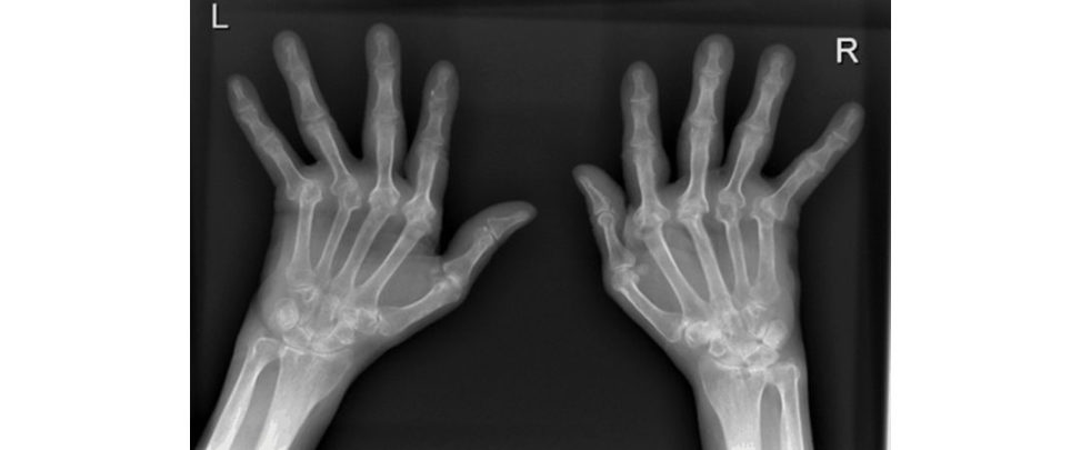
This is a posteroanterior X-ray of both hands showing a symmetrical polyarthropathy typical of a rheumatoid hand.
What other features are associated with rheumatoid hand?
swelling of the joints secondary to synovitis, muscle wasting and surgical scars.
What do you mean by a Swan-neck and Boutonnière’s deformity? [This is more likely to be a quick question in the clinicals rather than a detailed viva question.]
Hyperextension of the PIP joints with flexion of the DIP joints (Swan-neck deformity). Flexion of the PIP joints and hyperextension of the DIP joints (Boutounnière deformity). Factual rote-learned classification systems that do not necessarily help in planning management have fallen out of favour as viva questions.
What would be your management options in this patient?
I would also address the realistic expectations from operative intervention of the hands. If the patient is struggling with pain I would offer her MCP joint replacements using silastic implants. I would explain surgery aims to correct the ulnar deviation of the fingers improve any pain from those joints and the appearance of the hand, and possibly the ability to pinch (the range of movement and grip strength are unlikely to improve).
You have decided to put the patient on the waiting list for MCP joint replacement. Any challenges you could foresee in this case?
The challenges can be divided into pre-, intra- and postoperative. The challenges intra operatively include skin condition, soft -tissue release and correct balancing and osteopenic bone. The challenges postoperatively include wound healing, risk of infection and postoperative splinting and hand therapy.
What about medications in the rheumatoid arthritis patient?
However, newer biological factors such as etanercept and infliximab should be discontinued for 1 week and 6 weeks respectively, to reduce risk of infection postoperatively [2].
What are the complications of MCP joint replacement?
I would divide the complications in to early and late complications Early complications include joint infection stiffness, implant dislocation and incomplete correction of deformity.
Figure 17.6 Radiographs of hand demonstrating features of rheumatoid arthritis Erosions MC Pand PIP joints. Bilateral symmetrical involvement. Never the DIP joint. Destructive changes at the wrist. Look for periarticular os teopenia, uniform joint space loss, bone erosions and soft -tissues welling. Joint subluxation and subchondral cysts, ulnar deviation MCP joints.
References#
1. Grennan DM, Gray J, Loudon J, FearS. Methotrexate and early postoperative complications in patients with rheumatoid arthritis undergoing elective orthopaedic surgery. Ann Rheum Dis. 2001;60:214–217.
2. Scanzello CR, Figgie MP, Nestor BJ, Goodman SM. Peri operative management of medications used in the treatment of rheumatoid arthritis . HSS J. 2006;2(2):141–147.
3. MP joint arthroplasty. www.wheelessonline.com/ortho/mp_joint_arthroplasty
Structured oral examination question 7#
Thumb carpometacarpal (CMC) joint arthritis
Please describe the X-ray findings (Figure 17.7a).
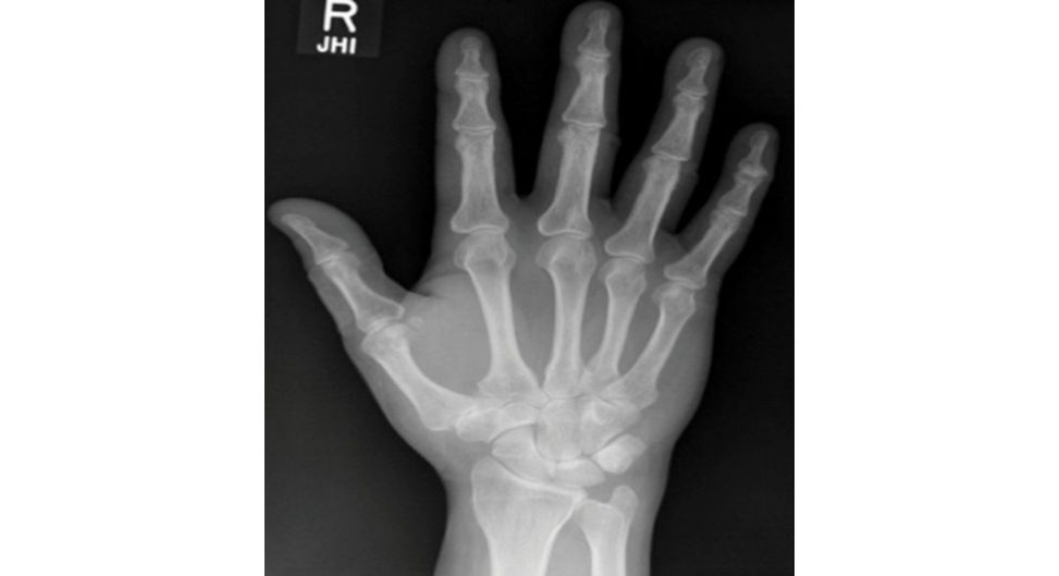
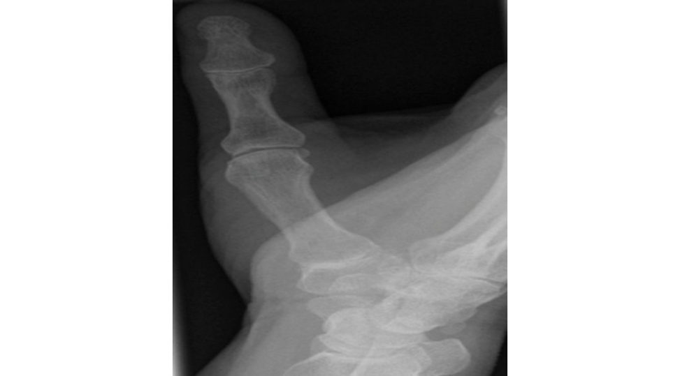
A Robert’s view, which is a true AP view of the thumb CMC joint, is taken with the elbow extended, the forearm fully pronated and the thumb abducted. This is shown in the X-ray below (Figure 17.7b).
Do you know any classification system for thumb CMC joint arthritis?
I would classify thumb CMC joint arthritis using the Ea ton and Litiler classification. Stage I: slight widening of the joint (2° effusion or ligament laxity). Stage II: slight joint space narrowing with sclerosis osteophytes < 2 mm, < 1/3 metacarpal base subluxation.
These X-rays belong to a 65-year-old lady with a painful thumb for the past 2 years. How would you proceed?
I would ask about hand dominance, night pain, effect on ADLs and treatment to date. Pain can be either diffuse or radial wrist pain up to the thumb MCP joint. Intersection syndrome (tender proximal to the styloid). Concomitant CTS (high volume of patient sin hand clinics have both CTS and basal thumb OA). The management of thumb CMC arthritis includes non-oper at ive and operative. Non-operativ e includes analgesia, activity modification, thumb splint and therapeutic injection of local anaesthesia into thumb CMC joint (as per NICE guidelines on osteoarthritis).
This quality standard covers diagnosing, assessing and managing osteoarthritis in adults.
What would YOU perform?
I would offer the patient simple trapeziectomy if all non-operativ e management has failed [1]. Prof. Davis et al. published their study in 2012 with a minimum 5 years follow-up of 153 thumbs with trapeziometacarpal osteoarthritis randomized into three groups to undergo either simple trapeziectomy, trapeziectomy with palmaris longus interposition or tr apeziectomy with ligament reconstruction and tendon interposition (LR TI) using 50% of the flexor carpi radialis tendon [2]. The study showed no benefit to tendon interposition or ligament reconstruction in the longer term compared to trapeziectomy alone in regard to thumb pain, function and strength. A Cochrane review in 2015 reported low-quality evidence to guide management, with insufficient evidence to suggest LRTI had additional benefit. LRTI patients experienced more adverse events.
Which ones?
I would explain preoperatively this is mainly a pain-relieving procedure, she would require hand splinting for 5–6 weeks postoperatively and is likely to have some loss of grip strength on a permanent basis.
What are the complications of tr apeziectomy?
Early – infection, haematoma formation, and injury to the superficial sensory nerves.
What would you offer if this is a 34-year-old male manual worker?
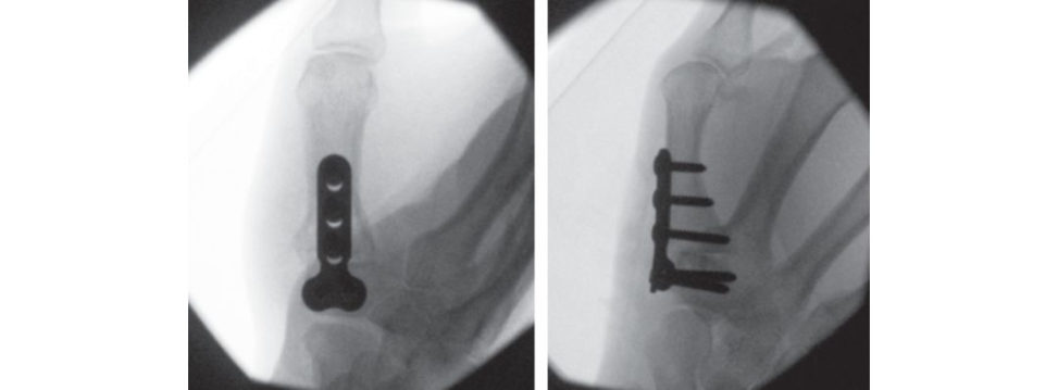
However, I would also counsel the patient that fusion has a higher incidence of complication such as non-union ( Figure 17.7c).
You mentioned the grind t est. Show me how you do it on my hand.
The testis positive if this causes pain and then the pain goes with rotation and distraction.
How would you try to address the adduction deformity of the thumb?
However, if there is ongoing deformity and unacceptable reduction in handspan following trapeziectomy, then I would consider an extension osteotomy of the thumb metacarpal.
How would you try to address the hyperextension at the MCP joint?
The hyperextension is greater than 30° and therefore if left untreated may cause a secondary swan-neck deformity of the thumb.
Figure 17.7a Posteroanterior (PA) radiograph, wrist.
Figure 17.7b Robert’s view (true AP view of the thumb CMC joint).
Figure 17.7c Radiographs demonstrating fusion CMC joint, as this possibly allows improved grip strength in a manual worker, but has a higher incidence of complications such as non-union.
References#
1. Davis TR, Brady O, Barton NJ, Lunn PG, Burke FD. Trapeziectomy alone, with tendon interposition or with ligament reconstruction? J Hand Surg Br. 1997;22(6):689–694.
2. Gangopadhyay S, McKenna H, Burke FD, Davis TR. Five- to 18-year follow-up for treatment of trapeziometacarpal osteoarthritis: a prospective comparison of excision, tendon interposition, and ligament reconstruction and tendon interposition . J Hand Surg Am. 2012;37(3):411–417.
Structured oral examination question 8#
Flexor tendon sheath infections
Please describe the clinical picture (Figure 17.8).
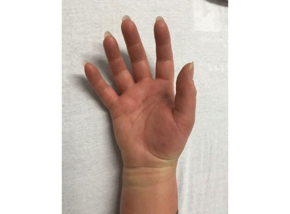
This clinical picture shows diffusely swollen and erythematous fingers, right hand. The fingers are held inflexion.
What’s your main concern and how would you approach this patient?
My main concern is flexor sheath tenosynovitis. I would examine the hand to look for any other wounds, collection and tenderness along the flexor sheath on palpation. I would also request a plain X-ray of the digit involved to exclude the presence of foreign bodies.
Who and how is this underlying condition classically described?
The classic description of flexor sheath tenosynovitis is by Kanavel [1]. P ain on passive extension of the digit was discussed in subsequent reports. Pain with passive stretching of the involved digit is perhaps the most sensitive test.
You have made the diagnosis of flexor sheath tenosynovitis. How would you manage this patient?
I would place my proximal incision over the A1 pulley at the distal palmar crease with complete release of this structure to gain access to the flexor sheath. A second oblique incision is made over the region of the A5 pulley, taking care to avoid injury to the neurovascular bundle. The sheath is copiously irrigated with a minimum of 500 ml normal saline but avoiding excessive fluid extravasation in to the digit because this can result in necrosis of the digit. Postoperatively, the hand should be splinted, elevated, discussion with the microbiologist regarding appropriate intravenous antibiotics continued for 5–7 days until the redness and inflammation has subsided.
Is there any role incontinuous catheter irrigation postoperatively?
A retrospective review comparing the intraoperative incision, drainage and irrigation with or without.
What are the complications if this condition is not treated promptly?
The complications include early skin loss tendon necrosis and rupture, tendon adhesions, deep palmar space infection, septic arthritis, osteomyelitis and hand stiffness.
What about an open approach for flexor sheath irrigation for acute pyogenic flexor tenosynovitis?
The approach uses longitudinal midaxial or a volar zig-zag Brunner’s incision with direct drainage of any purulent material. The midaxial approach is possibly preferable as there isless concern about skin coverage postoperatively. There is an increased risk of tendon necrosis, increased scarring and stiffness and significant morbidity.
When would you use an open approach?
The reis some evidence to recommend open treatment of stage III infections necrosis of the tendon, pulleys or tendon sheath).
You have mentioned stage 3 infection.
Stage 2: purulent fluid, granulomatous synovium. Stage 3: necrosis of the tendon, pulleys or tendon sheath.
Figure 17.8 Flexor sheath tenosynovitis. The reis diffuse swelling of the fingers. The fingers are flexed.
References#
1. Kana velA. Infections of the Hand: A Guide to the Surgical Treatment of Acute and Chronic Suppurative
Processes in the Fingers. London: Balliere, Tindal land Cox; 1939.
2. Lille S, Hayakawa T, Neumeister MW, et al. Continuous postoperative catheter irrigation is not necessary for the treatment of suppurative flexor tenosynovitis . J Hand Surg (Br). 2000;25(3):304–307.
Structured oral examination question 9#
Flexor tendon injury
Can you describe this clinical photograph and what it represents (Figure 17.9)?
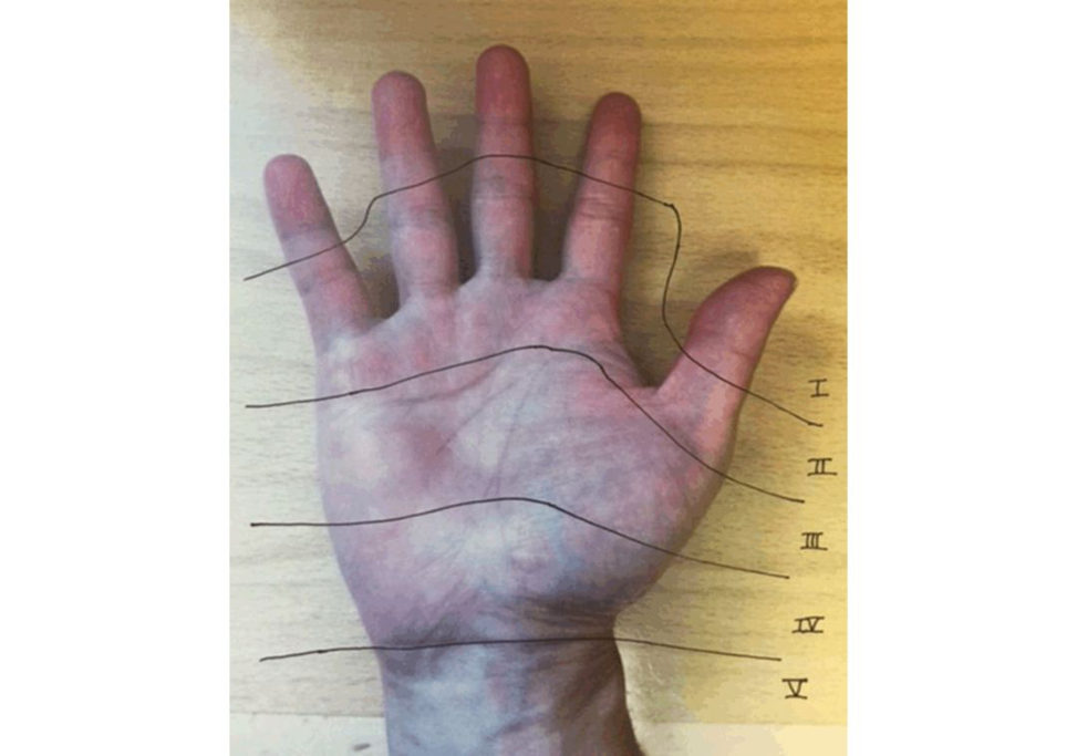
Zone 1 – flexor digitorum profundus distal to insertion of flexor digitorum superficialis. Zone 2 – insertion of flexor digitorum superficialis to proximal edge of A1 pulley (no man’s land). Zone 4 – within the carpal tunnel.
When would you consider repairing a lacerated tendon?
Studies have shown that a lacerated tendon with less than 60% cross-sectional area involvement can be managed without repair, but with debridement and early mobilization.
What is the goal of tendon repair and how would you perform it?
The goal of tendon repair is to maintain sufficient strength to avoid rupture with either passive or active movement during mobilization. Circumferential epitenon suture has also been shown to significantly increase the resistance of the tendon repair to gap formation aswell as adding 20% to the ultimate tensile strength of the repair [2].
What is the significance of Zone 2 flexor tendon injury?
Bunnell referred to this area as no man’s land because the initial results of the tendon here were so poor. The advances in suture techniques, better understanding of the tendon morphology and its biomechanics as well as early active mobilization rehabilitation protocols have resulted in better outcomes in Zone 2 flexor tendon repair.
What is the postoperative rehabilitation following flexor tendon repair?
Postoperative rehabilitation aims to mobilize early (48 hours postoperatively) to prevent adhesions. Most patients will be put into an active place and hold’ regimen rather than traditional passive range of motion regimen. This was supported by a randomized controlled trial by Trumble et al. in 2010 which showed that active motion had significantly fewer flexion contractures, better satisfaction scores and improved range of motion than passively rehabilitated patients Importantly, there was no difference in the tendon re-rupture rate [3].
Figure 17.9 Flexor tendon injury zones.
References#
1. Bishop AT, Cooney WP 3rd, Wood MB. Treatment of partial flexor tendon lacerations: the effect of tenorrhaphy and early protected mobilisation . J Trauma. 1986;26(4):301–312.
2. Pruift DL, Manske PR, Fink B. Cyclic stress analysis of flexor tendon repair. J Hand Surg (Am).
1991;16(4):701–707.
3. Trumble TE, Vedder NB, Seiler JG 3rd, et al. Zone-II flexor tendon repair: a randomised prospective trial of active place-and-hold therapy compared with passive motion therapy. J Bone Joint Surg (Am).
2013;38:1800–1802.
Structured oral examination question 10#
Enchondroma
Please describe the clinical picture of a 10-year-old girl who fell onto her left hand a few hours ago (Figure 17.10a).
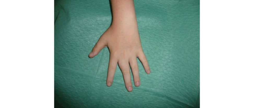
There is widening of the interspace between the index and middle fingers.
How would you manage this patient?
I would provide adequate analgesia for the patient and arrange an X-ray of the middle finger to exclude fracture.
Please describe the X-ray of the left middle finger of this patient Figure 17.10b).
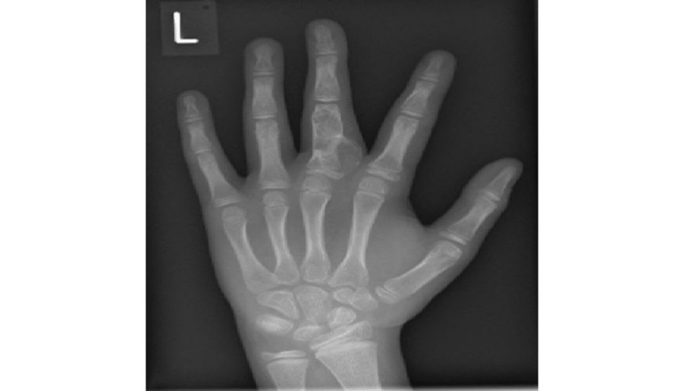
This is a posteroanterior (PA) view of the left hand of a skeletally immature patient The radiograph shows a pathological fracture through the cystic lesion affecting the proximal phalanx of the middle finger. I would confirm this further with lateral and oblique views of the middle finger.
How would you manage this fracture?
As discussed earlier, I would take a detailed history and examination of the affected finger. History includes rapid increase in size or pain over the middle finger. I would manage this fracture conservatively with buddy strapping for 2 weeks provided there is no rotational deformity. I would review them at 6 weeks and then annually. If it continued to increase in size, I would refer to a hand surgeon for partial excision and bone gratiing [ 1].
What are you going to tell the parent who is concerned regarding this lesion?
I would discuss and reassure the patient and parent that this is a benign cartilage tumour of bone that is mostly asymptomatic. The malignant risk with a single lesion is low but the risk increases to 20–30% for Ollier’s disease and near 100% for Mafucci’s disease.
Figure 17.10a Clinical picture of an 11-year-old’s hand.
Figure 17.10b Radiograph of the child’s hand.
References#
1. O’Connor MI, Bancroft LW. Benign and malignant cartilage tumours of the hand. Hand Clin.
2004;20(3):317–323.
2. Athanasian EA. Bone and soft tissue tumours. In DP Green, RN Hotchkiss, WC Pederson et al. (Eds.),
Green’s Operative Hand Surgery, 5th ed. Philadelphia: Churchill Livingstone; 2005: pp. 2211–2263.
Structured oral examination question 11#
Wrist ganglion
Please describe this clinical picture (Figure 17.11a).
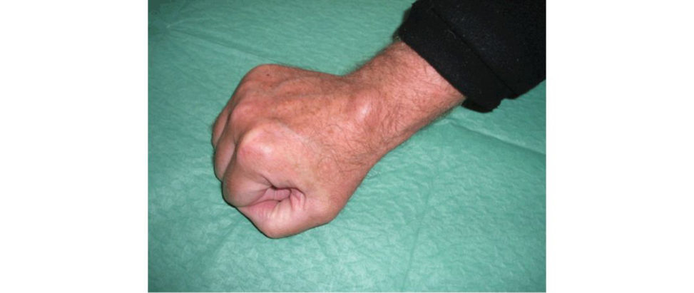
This clinical picture of a clenched right fist shows a swelling over the dorsoradial aspect of the wrist, suggestive of ganglion.
How would you confirm your diagnosis?
I would request radiological investigations such as ultrasound or magnetic resonance imaging (MR Ito confirm my diagnosis.
This is the investigation that has been performed. Please comment (Figure 17.11b).
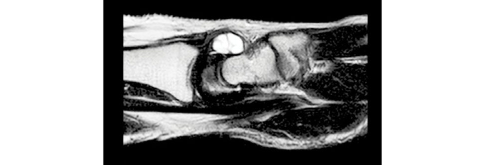
The MR scan confirms the diagnosis of ganglion that is likely to have arisen from the scapholunate ligament.
What is the aetiology of this condition?
Ganglion is a mucin-filled synovial cyst with no true epithelial lining that arises from the tendon sheath or joint capsule.
How would you manage this?
If there is pain affecting function, especially with forced wrist extension, then aspiration or surgical excision may be offered.
What complications can occur with aspiration?
Haematoma, bruising, infection and reoccurrence.
What is the incidence of reoccurrence following aspiration?
The reoccurrence rate following aspiration is 50%.
What would you do if the ganglion reoccurs after aspiration?
If the ganglion reoccurs after aspiration and the patient remains symptomatic, I would offer the patient surgical excision, taking into consideration the reoccurrence rate of 5%.
What complications can occur with surgical excision?
Haematoma, bruising, infection neurovascular damage and reoccurrence.
What is the second most common hand swelling?
A giant cell tumour or xanthelasma.
Correct.
Figure 17.11a Clinical picture of wrist.
Figure 17.11b Imaging of the same wrist.
Structured oral examination question 12#
Dupuytren’s disease
Please describe the clinical photograph of a 45-year-old with this problem in the non- dominant hand (Figure 17.12a).
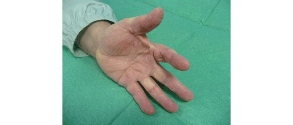
The clinical picture shows a thick cord crossing the first webspace of the left hand causing a web space contracture, and fixed flexion at the metacarpophalangeal (MC Pand proximal interphalangeal (PIP) joint of index finger. The cord seen is known as the commissural band, usually associated with Dupuytren’s diathesis. The degree of diathesis is considered important in predicting recurrence and extension of Dupuytren’s disease after surgical management [1].
What is the primary cell involved?
They are probably derived from fibroblasts and contain smooth muscle actin which leads to contracture of the cord.
What other risk factors are associated with this disease?
Other risk factors for Dupuytren’s disease include diabetes, epilepsy, high alcohol intake,
What structures make up a spiral cord and how does it affect the neurovascular bundle?
The pretendinous, lateral and spiral bands and Grayson’s ligament make up the spiral cord.
How do you classify the disease?
The British Society for Surgery of the Hand (BSSH) classifies Dupuytren’s disease as: Mild – no functional problems, no contracture or metacarpophalangeal joint contracture of less than 30°.
What are the management options?
Non-operativ e management includes observation ± night splintage, and collagenase injections.
Operative intervention includes fasciotomy (division of the cord), fasciectomy (excision of the cord)
and dermofasciectomy (cord and overlying skin excised) and skin gratiing.
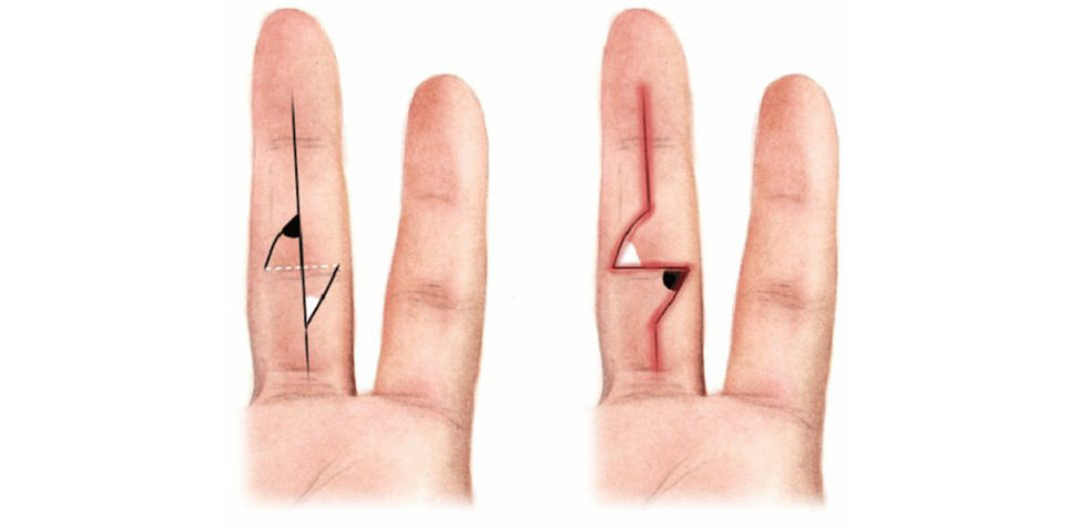
What is a Z-plasty? Can you draw it?
Angles should be made at 60° to the incision to achieve a 75% increase in length (Figure 17.12b).
How would you consent for a fasciectomy?
You will beseen at 10 days for removal of your sutures. Complications include the following [2]: Early infection, bleeding and haematoma formation, arterial or nerve injury, necrosis of digit leading to amputation tendon injury and delayed wound healing. Late joint stiffness recurrence (50% recur but most do not require further surgery) and reoperation incomplete correction of deformity and complex regional pain syndrome.
Do you know any new treatments for Dupuytren’s contracture?
Two randomized controlled studies of 374 patients comparing Xiapex to placebo have shown benefit with 60% showing correction to 5° of full extension [3]. The technique is appropriate treatment for apa tien t with single-digit involvement and contractures limited to the palm and MCP joint. The patient can receive up to a maximum of three injections per c ord, limited to two digits (one at a time) a t 4-week intervals.
Figure 17.12a Clinical picture of a hand.
Figure 17.12b How to draw a Z-plasty. 1. Draw perpendicular (whited oft ed line) to longitudinal incision. 2. Mark flaps (white and black angles are at 60°). 3. Crossover flaps as shown.
References#
1. Hindocha S, Stanley JK, WatsonS Bay atA. Dupuytren’s diathesis revisited: evaluation of prognostic indicators for risk of disease recurrence. J Hand Surg Am. 2006;31(10):1626–1634.
2. Hayton MJ, Gray ICM. Dupuytren’s contracture: a review. Curr Orthop. 2003;17:1–7.
3. Hurst LC, Badalamente MA, Hentz VR, et al. Injectable collagenase Clostridium histolyticum for
Dupuytren’s contracture. N Engl J Med. 2009;361(10):968–979.
Structured oral examination question 13#
Kienbock’s disease
Please describe this X-ray (Figure 17.13a).
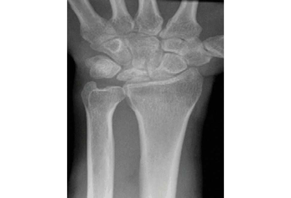
There are no radiological arthritic changes to the surrounding joints. The radiograph is suggestive of Kienbock’s disease (avascular necrosis of the lunate).
What are the predisposing factors for Kienbock’s disease?
Predisposing factors include ulnar minus variant – this is thought to lead to increased.
What are the patterns of intraosseous blood supply for lunate?
Gelbermann described the various patterns of intraosseous blood supply for lunate – Y pattern (60%), I pattern (30%) and an X pattern (10%) – in his paper in 1980. Single or repetiv e microfractures can result in disruption of the blood supply to the lunate. As such, apa tien t with I-pattern lunate blood supply is more susceptible to AVN.
What classification do you know for Kienbock’s disease?
The stages as described by Lichtman are as follows: Stage I – plain radiographs normal (changes seen on MRI). Stage II – lunate sclerosis seen on plain radiographs. Stage IIIA – fragmentation and collapse of lunate without fixed scaphoid rotation derangements or instability). Stage IIIB – fragmentation and collapse of lunate with fixed scaphoid rotation, decreased carpal height and ulnar migration of the trique trium. Stage IV – radiocarpal and midcarpal arthritis, degenerative wrist changes.
What is the role of MRI in Kienbock’s disease?
MRI will show decreased signals on T1- and
T2-weighted images.
EIXAMINER : What’s the differential diagnosis?
CAINDIDATE : The differential diagnosis includes pos t-traumatic arthritis; fracture; carpal instability.
What are the management options for Kienbock’s disease?
However, in symptomatic patients with Stage I/II/IIIA disease and if ulnar minus, I would offer a joint levelling procedure, either a radial shortening orless commonly an ulnar lengthening. Other options are a vascularized bone graft with 4, 5-ICSRA , core decompression of the distal radius or a distal radial osteotomy. Surgical options for Stage IIIB/IV disease include a neurectomy, or salvage procedures such as a proximal row carpectomy or a wrist arthrodesis.
How would you manage this case?
If these were, the wrist appears to be ulnar minus and I would offer a joint levelling procedure with a radial shortening using a volar locking plate (Figure 17.13b).
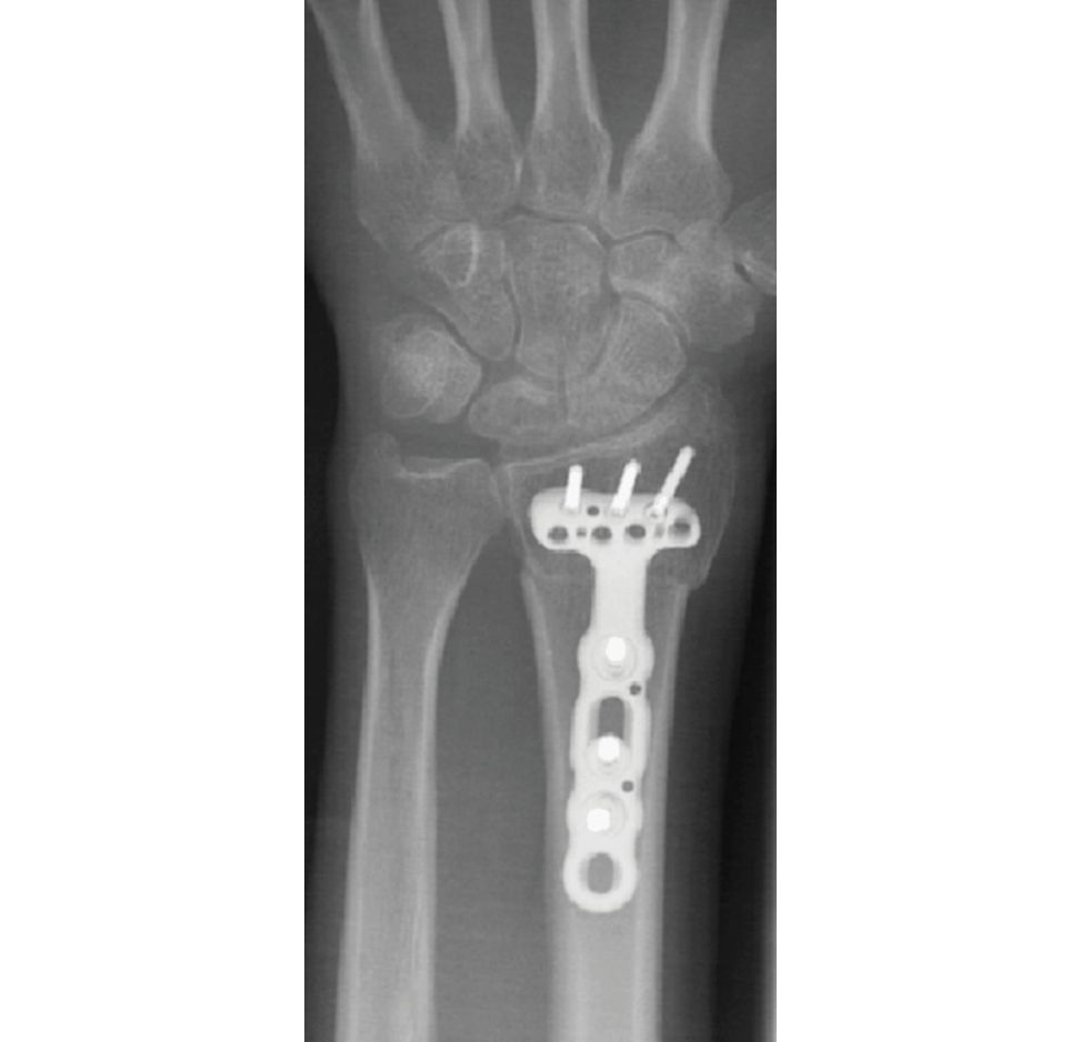
Figure 17.13a Posteroanterior (PA) radiograph of left hand.
Figure 17.13b Posteroanterior (PA) radiograph demonstrating a joint levelling procedure (radial shortening).
Reference#
1. Gelberman RH, Bauman TD, Menon JAke son WH. The vascularity of the lunate bone and Kienbock’s disease. J Hand Surg Am. 1980;5(3):272–278.
Structured oral examination question 14#
Ulnar collateral ligament (UCL) injury of thumb
What testis being done here (Figure 17.14a)?
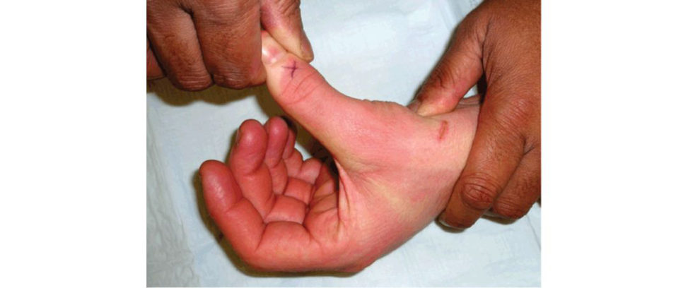
The testis done by stabilizing the thumb metacarpal and applying a valgus stress to the thumb MCP joint with the thumb flexed at 30°.
Tell me about ulnar collateral ligament injury. What is a Stener lesion and its significance?
Acute ulnar collateral ligament injury is known as skier’s thumb and chronic injury is known as a gamekeeper’s thumb. A St ener lesion requires surgical intervention.
How would you differentiate between complete and incomplete tear?
Instability of the MCPJ inflexion suggests an isolated proper UCL injury whereas instability inextension suggests the accessory ligament is involved. Plain radiographs would not demonstrate any joint widening on the ulnar border of the MCPJ in an incomplete lesion.
When would you decide to operate on UCL injury?
I would opt to operate if there’s more than 20° of opening on radial stressing of an acute UCL injury or the presence of a Stener lesion.
How does a chronic UCL injury present?
Patients will complain of pain mainly on the inner side of the thumb when stressing the thumb with activitie slik e gripping or pinching.
How would you manage a chronic UCL injury?
In a symptomatic chronic UCL injury, this would need reconstruction using the palmaris longus tendon as a graft.
Figure 17.14a Clinical picture of stress testing ulnar collateral ligament (UCL).
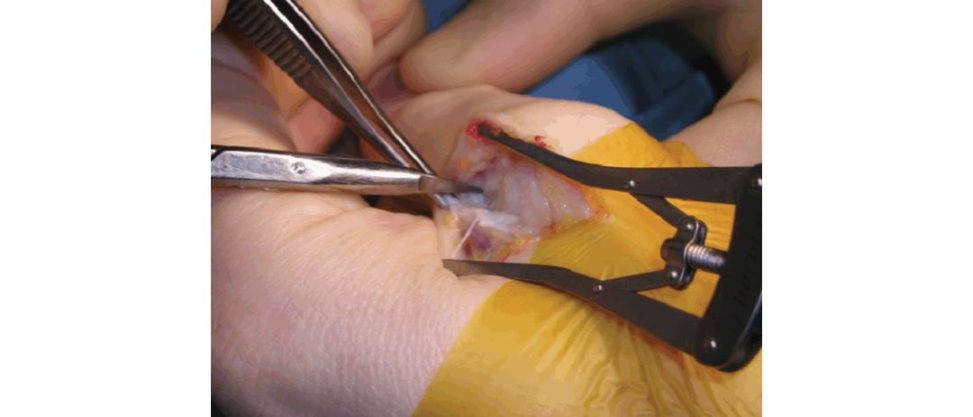
Figure 17.14b Intraoperative picture showing an approach to the medial side of the thumb MCP joint for an ulnar collateral ligament repair. The scissor tips are pointing a t the bare insertion of the ligament to the base of the proximal phalanx. This is a chronic case and the scarred proximal ligament is lying bunched up over the metacarpal head.
Structured oral examination question 15#
Boutonnière deformity
A rugby player has injured his finger during a game. He has pain and swelling and attends casualty where radiographs are taken. Describe what you see and the likely diagnosis (Figure 17.15a).
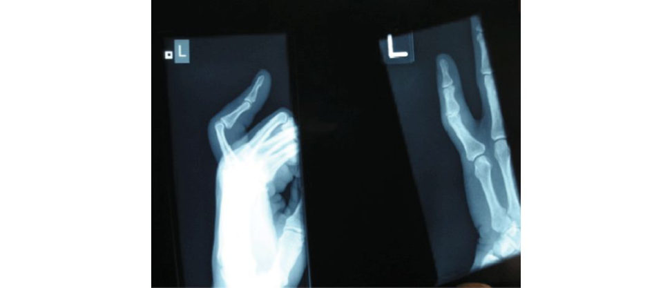
The radiographs show flexion at the PIP joint of the litile finger and hyperextension at the DIP.
How would you manage this?
I would test the central slip of the extensor mechanism by Elson’s test – the PIP joint is flexed to 90° over the edge of a table, and the patient is asked to extend the finger against resistance (the examiner presses on the middle phalanx). I would offer a dynamic splint – a Capener – to keep the PIP joint passively extended but allow active flexion (Figure 17.15b).
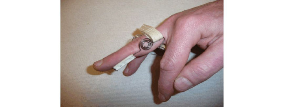
Figure 17.15a Anteroposterior (AP) and lateral radiographs of alit ile finger.
Figure 17.15b Picture of a splint used to manage a Boutonnière deformity.
Structured oral examination question 16#
Swan-neck deformity
These are clinical pictures of a 60-year-old lady complaining of difficulty making a full fist with her left hand ( Figure 17.16).
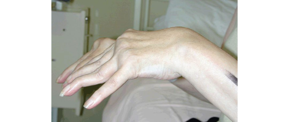
The photograph shows hyperextension at the proximal interphalangeal joint (PIPJ) and.
What deformity is this and what is the pathophysiology of it?
Candidate should have followed through and said the picture was highly suggestive of swan- neck deformity in keeping with a picture of a generalized symmetrical polyarthropathy, most likely rheumatoid arthritis.
What is the classification system associated with this deformity?
Swan-neck deformity in a rheumatoid hand is classified according to PIPJ mobility and radiographic appearances: Type I – PIP joint flexible in all positions.
What are the causes of this deformity?
The primary pathology is a lax volar plate; this may be caused by rheumatoid arthritis trauma or generalized ligamentous laxity. This may occur at different sites such as at MCP, PIP or DIP joints. PIP joint FDS rupture with unopposed PIPJ extension resulting in intrinsic contracture. The excursion of the lateral bands is restricted; therefore, the extension force is not transmift ed to the terminal tendon. The DIP joint extension force is transferred to the PIPJ central slip.
What are the surgical treatments for swan-neck deformity?
The patient hand function and expectations must be fully assessed. I would divide the surgical procedures according to the joints involved such as below: MCP joint problems can be addressed with synovectomy and MCP joint arthroplasty. The bandis passed volar to Cleland’s ligament and fixed to itself around the A2 pulley attachment (as for FDS tenodesis). PIP joint problems, FDS tenodesis can be performed with a single slip of the FDS being passed around the A2 pulley sewn to itself holding the PIP joint flexed. The extensor expansion including lateral bands and collateral ligaments is released with removal of osteophytes and with a synovectomy if required, ensuring full PIP joint range of movement. DIP joint problems can be addressed with a DIP joint fusion or possibly a dermodesis.
What are the complications of surgery?
I would divide the complications of surgery into those related to the actual surgical procedures,
Figure 17.16 Swan-neck deformity, fingers.
Complications related to surgery
Soft -tissue procedure infection, failure to correct deformity, overcorrection of deformity,
Arthrodesis procedure non-union, malunion, implant failure or prominence.
fracture, dislocation, loosening, stiffness, tendon/ligament damage,
Complications related to medications
Medications should be managed in conjunction with a rheumatologist. Coverage should be provided if the patient has been on 10 mg of prednisolone per day for 1 week or more within the last 6 months.
Aspirin/NSAIDs use affects platelet function and bleeding during and after surgery.
Anft-TNF and other biological medications may affect wound healing and increase the risk of infection postoperatively. Speak to a rheumatologist preoperatively.
Complications related to systemic diseases
Atlantoaxial subluxation or basal invagination is a possible complication of RA and the C-spine anaesthetist should make a full assessment preoperatively.
Cardiac disease mostly not related toRA, conduction blocks secondary to granulomas or nodules,
Airway disease along with cervical spine pathology,
Lung disease: pleural effusion; More severe lung disease such as interstial pneumonitis; bronchiolitis can affect operative risk.
These may be secondary to rheumatic drug therapy such as gold, penicillamine or methotrexate.
Bone disease can lead to osteoporosis and care should betaken when handling these patients.
Vasculitis can lead to non-healing ulcers and neuropathy.
Renal disease: the kidneys may be affected by vasculitis or amyloidosis (chronic disease).
Pharmacological treatments may also damage the kidneys.
Haematological RA patient may have anaemia or neutropenia (Felty’s syndrome).
Infection patients with RA may have an increased susceptibility to infection postoperatively.