Chapter 24 Musculoskeletal oncology
Definitions#
As in all other areas of the viva examinations, clear thought process, and are in command of the subject matter.
Neoplasm/tumour:
A growth or swelling, which enlarges by cellular proliferation more rapidly than surrounding normal tissue and continues to enlarge after the initiating stimuli cease.
Malignant tumour:
and to distant metastasis usually via the vascular or lymphatic systems.
Benign tumour:
Benign tumours do not metastasize but can still exhibit locally aggressive behaviour.
Sarcoma:
A diverse and rare group of malignant tumours of mesenchymal/connectiv e tissue origin.
Tumours of peripheral nerves are often included in this group.
Generic structured oral examination question 1#
Biopsy
So how would you obtain a tissue diagnosis?
In general terms this can be performed by excisional, incisional or percutaneous means, but I would not perform a biopsy without first having discussed the case with a bone and soft tissue tumour MD T.
Good. Let’s suppose you are the bone tumour surgeon now. When might you perform an excision biopsy?
Hence, this type of biopsy is really only applicable to benign lesions where the imaging has been diagnostic, for example lipomas, or where the lesion is small and superficial such that excision biopsy would not compromise later re-excision.
OK, tell me how you would perform an incisional biopsy.
I would plan the incision using the imaging and position it such that the entire biopsy tract could be excised enbloc during the definitive resection, and such that it does not contaminate more than one compartment or key neurovascular structures.
We perform most of our biopsies percutaneously now. Do you know any advantages or disadvantages to doing it this way?
I’ve seen biopsy performed by Tru-Cut needle. W elker et al. have shown that it is safe, has a low complication rate and reliably provides enough tissue for diagnosis and treatment planning [1]. The disadvantage is that necrosis and mitotic rate are less reliable on core needle, but this rarely affects management, and an incisional biopsy can always be performed subsequently if more information is required.
What do you understand by a marginal margin?
A marginal margin, as described by Enneking, is when the resection line passes through the reactive zone of the tumour being excised [2].
Explain to me what you mean by the reactive zone.
This is the reactive zone. Hence, if a resection line passes through this reactive zone, as in a marginal margin, then micronodules of tumour are likely to be left behind, increasing the risk of a local recurrence.
So, what other margins did Enneking describe and what do you understand by them?
Enneking described three other possible margins. In wide margins it is still possible that tumour will remain in the form of skip lesions. Finally, he described the radical margin, where the entire compartment in which the tumour resides is excised enbloc, in theory removing the entire tumour [2].
Generic structured oral examination question 2#
Staging
So what stage is this tumour?
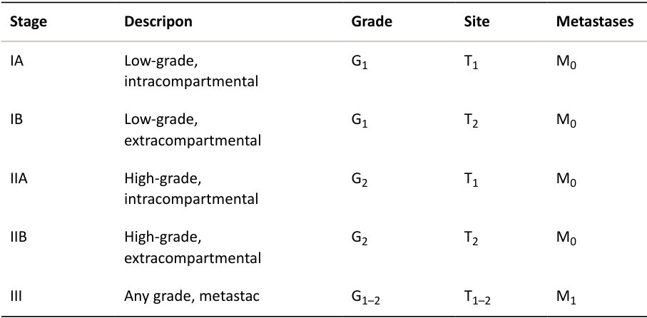
I would stage this tumour using the Musculoskeletal Tumour Society staging system as described by Enneking [3]. The Enneking system is the easiest to remember and can be applied equally to bony and soft -tissue sarcomas. The other commonly used system is the American Joint Commift ee on Cancer (AJCC) system, which is more complicated. The AJCC also has separate systems for bony and soft -tissue tumours. Table 24.1 Enneking/MSTS staging system [4].
Structured oral examination question 1#
Osteochondroma
This young lad has been referred to you urgently by his GP after his mum brought him in with a firm lump on the front of his left thighT ell me about his X-ray (Figure 24.1).
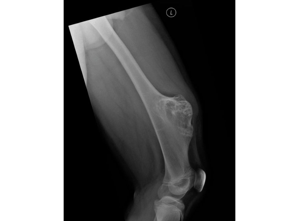
Figure 24.1 Osteochondroma.
This is a lateral radiograph of his left femur including the knee joint but not the hip joint.
What makes you think it’s an osteochondroma?
Well, the cortices are incontinuity with the bone as is the medullary cavity, and the lesion is extending out from the metaphyseal region of the distal femur, which is the most common site for these (25%).
OK, so you get an MRI, which shows a nice thin cartilage cap and no worrying features. How are you going to treat it?
First I’d take a history and examine the child. I’d want to know if it is tender or symptomatic before I decide what to do.
It’s not tender and it only bothers him occasionally if he knocks it, but his mother is adamant she wants it removed.
There is also a chance of fracture during the operation and afterwards as it’s a large sessile lesion and removing it will weaken the anterior cortex of the femur considerably.
His mum still w ants it removed and she’s worried that it’s going to become cancer.
If this is a solitary lesion then malignant change is very rare indeed (< 1%). The textbooks often quote figures of 10% but it is probably more like 1–5%. Examiners may show an example of a solitary osteochondroma in an area that is General advice: difficult to access for the purposes of excision, but then insist that the patient wants it removed, e.g. posterior, proximal tibia. A major consideration in determining the malignant potential of an osteochondroma is the thickness of its cartilage cap with thicknesses greater than 1 cm considered worrisome.
Structured oral examination question 2#
Enchondroma
Tell me about these radiographs of this chap’s right foot (Figure 24.2).
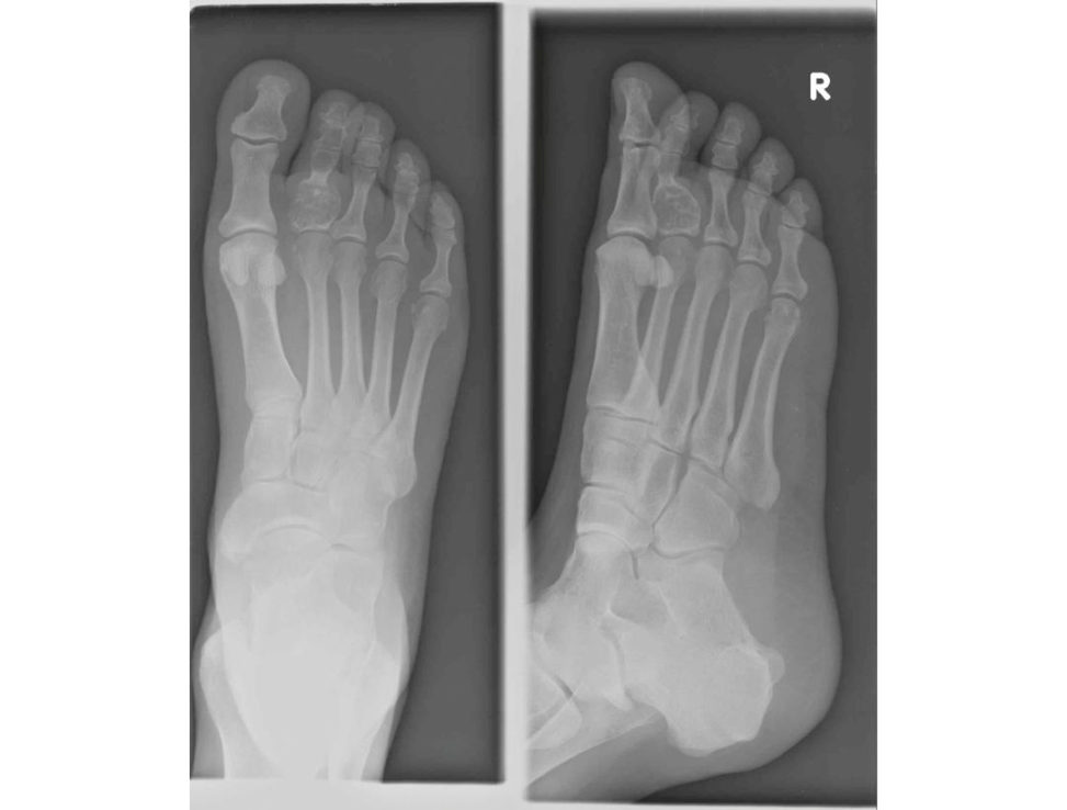
Figure 24.2 Enchondroma.
Well they’re AP and oblique views and they show an expansile, lytic lesion in the proximal phalanx of his second toe.
What do you think it is?
There’s also some stippled calcification within the substance of the lesion, which suggests a chondroid matrix.
How would you treat this lesion?
Well I would want to get more information, so I would take a full history and examination. Always work through history, examination and imaging You will never be criticized for discussing the diagnosis with a bone tumour MDT, but you will end up in a very tricky discussion with the examiners and fail if you have made the wrong diagnosis, it turns out to be malignant, and you’ve not discussed it with an MDT first.
Other points
50% of solitary enchondromas arise in the hands.
Malignant transformation is very rare, but when it does occur it is usually in large lesions of long bones.
Enchondromatosis = Ollier’s disease (risk of bone malignancy is 10%.
Enchondromatosis + haemangiomas = Maffucci’s syndrome (risk of malignancy reported anywhere from 25% to 100%).
Structured oral examination question 3#
Non-ossifying fibroma
Tell me about this radiograph (Figure 24.3).
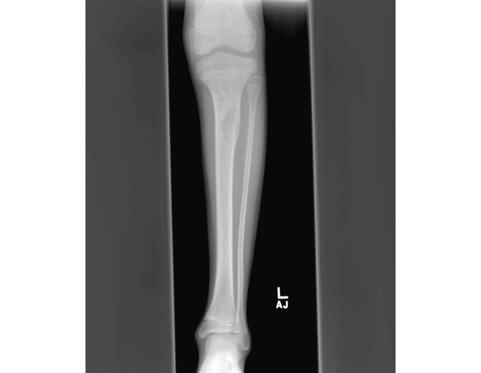
Figure 24.3 Non-ossifying fibroma.
This is an AP radiograph of a left lo wer leg of a child, which includes both the ankle joint and the knee joint. These features are typical of a non- ossifying fibroma.
Good. What else can you tell me about this lesion?
Non-ossifying fibromas are developmental or hamartomatous lesions. They are actually very common, and some have suggested an incidence of up to 35% in normal children.
How would you treat this lesion?
I can’t see any evidence of fracture. That being the case, this can be treated with observation only as these lesions normally resolve by adulthood. I would plan to keep the patient under review with surveillance radiography.
Again, you will not be criticize dif you say that would take advice from the bone tumour MDT.
Structured oral examination question 4#
Chondrosarcoma
This 60-year-old lady presented with pain and swelling around her lower back. What can you see on this CT scan (Figure 24.4)?
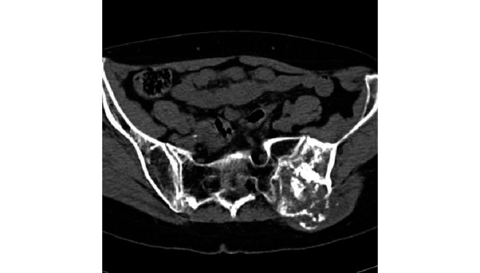
Figure 24.4 Chondrosarcoma.
This is an axial section showing the sacrum and iliac wings. The lesion has both ly tic and sclerotic elements to it.
What do you think the diagnosis might be?
Primary bone tumours can be classified according to their matrix as either bone-producing, cartilag e- producing, fibrous tissue-pr oducing, or non-matrix-producing. The patchy sclerosis within this lesion is in keeping with either a bone- or cartilag e-producing primary tumour, although I would not rule out other diagnoses without further investigations [ 5].
You’re right to suggesta primary lesion in this case. You’ve suggested bone- or cartilag e- producing as the likely matrix. Which do you think is more likely here?
It is most likely to be a chondrosarcoma. Only around 5% of osteosarcomas occur in the pelvis, whereas up to 30% of chondrosarcomas are pelvic in origin. It’s not the clearest image, but I’m trying to convince myself that there’s stippled calcification, which would indicate a chondroid lesion.
Very good. What treatment options are there for an aggressive-looking chondrosarcoma like this is?
so the only treatment option is wide local excision plus or minus reconstruction However, despite surgical excision.
So, what do you think the prognosis is for this high-grade lesion?
High grade III lesions, as you’ve intimated this one is, are metastatic ino ver 70% of cases and have only a 30% 5-year survival.
Structured oral examination question 3#
Chondrosarcoma
This is a very fit and well 50-year-old chap, who has come into Accident & Emergency after falling down the stairs at home, sustaining this injury to his left leg. T ell me how you are going to manage this (Figure 24.5).
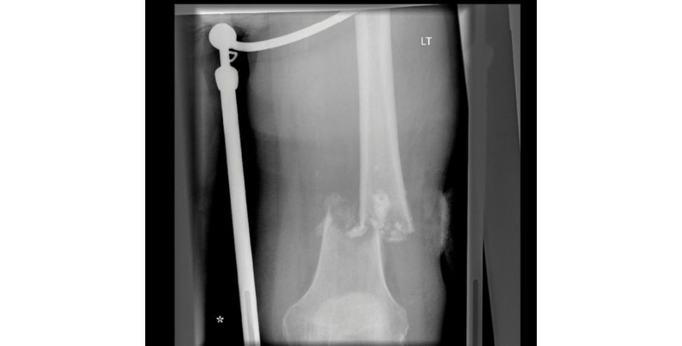
I would manage this patient initially using the principles of A TLS.
Fine. No issues with ABC and the patient is alert and orientated.
Moving on, I want to assess whether the patient has any other injuries, whether the limbis neurovascularly intact.
OK. This is his only injury. It’s an open fracture with a 1-cm wound on the lateral thigh. The limbis neurovascularly intact. How are you going to manage this?
If it’s an open injury then I would take a picture of the wound and cover it with a sterile- soaked swab. In this case I can see that a Thomas splint has been applied.
OK. So, shall I book this patient for theatre with a plan to perform a debridement of the wound and nailing of the fracture?
There’s some odd calcification within the medullary cavity, so I’m worried that this is a pathological fracture through a bony lesion.
Why does that make a difference?
It’s a rare situation, but if this is a pathological fracture through a bone tumour, and we open up the fracture site.
But doesn’t the open fracture need washing out?
It’s a small puncture wound, and I’ve put the patient on IV antibiotics, so I think the infection risk is low. You will never be criticized for taking advice, but you will fail if you have blazed on with treatment and taken this patient to theatre for washout and nailing. Always take timet o look carefully before answering, especially if a question on a fracture comes up in the adult and pathology viva station!
Figure 24.5 Chondrosarcoma 2.
Other points
Chondrosarcomas are the second most frequent primary malignant tumour of bone.
Chondrosarcomas are chemo- and radioresistant because of the presence of hyaline dense extracellular matrix, low mitotic activity and poor vascularity.
Low-grade (Gd1) tumours can be managed, extensive intralesional curettage, adjuvant therapy such as cryotherapy phenolization or ar gon beam laser followed by a void-filling procedure.
High-grade lesions invariably require en-bloc resection.
Phase II study using Imatinib).
Structured oral examination question 6#
Osteosarcoma [6]
This young lad presented with a painful knee and a lump after a football injury. What do you think of the X-ray (Figure 24.6)?
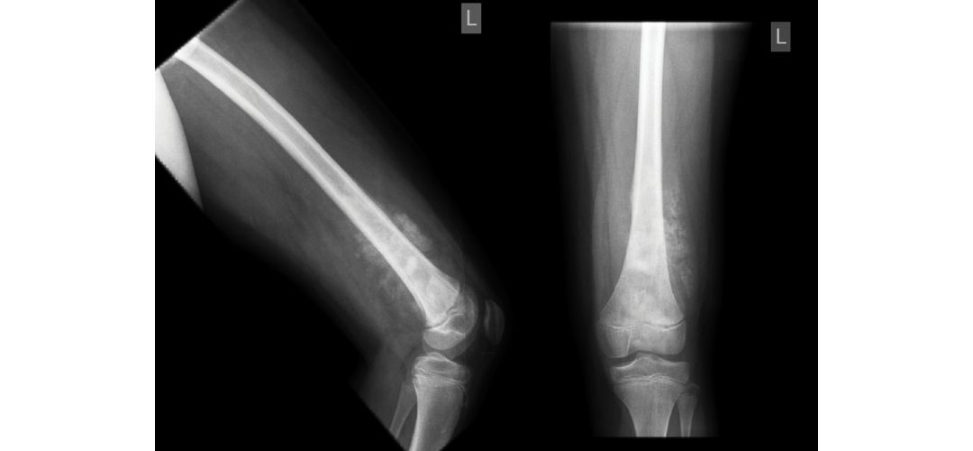
The reis an intramedullary sclerotic lesion with a wide z one of transition and the reis extension through the cortices and in to the soft tissu esTher e is sunray spiculation, butat this resolution I c an’t see an obvious Codman’s triangle.
What’s a Codman’s triangle?
I knew what a Codman’s triangle was, but I did not have a clear definition a t my fingertips (a triangle of reactive bone at the edge of the tumour where the periosteum is elevated).
So, what do you think the diagnosis is?
It is also in a classical position in the metaphyseal region of the distal femur, where about 35% of these tumours occur.
So how would you investigate it further?
I would refer the child on to a bone tumour MDT immediately rather than delay the process by organizing more investigations locally.
OK, so you’re working for the bone tumour MDT, what further investigations would you request?
I would request investigations to further delineate the tumour itself and I would arrange tests to assess for metastatic disease. These investigations can also be used to plan a biopsy. To stage the tumour one might initially get a chest X-ray, but CT scan of the chest is mandatory to look for metastases and these are sadly found in about 30% at diagnosis.
Tell me about the general principles of treatment in cases like this.
Before commencing treatment, a confirmatory tissue diagnosis is made by biopsy and staging investigations are completed.
neoadjuvant chemotherapy, imaging to look for recurrent disease or distant metastases.
Why does the treatment start with neoadjuvant chemotherapy? Why don’t we start by excising the tumour and then start chemotherapy?
First, to treat occult micrometastases, which are likely to be present in a much greater proportion of patients than the 30% who present with radiologically detectable metastases at diagnosis; second, to reduce the inflammation around the primary tumour, aiding later surgical resection; and finally , to allow assessment of response to the neoadjuvant chemotherapy, determine prognosis, and direct adjuvant chemotherapy.
You mentioned assessment of response to neoadjuvant chemotherapy. Why is this important?
Response of the tumour to chemotherapy treatment is measured as a percentage necrosis on histology of the resected specimen. A greater than 90% necrosis is considered a good response, and this carries a better prognosis than poor or non-responders.
Do you know of any novel treatments?
In a randomized trial, Meyers et al. showed that, when MTP was added to the standard chemotherapy regime of cisplatin, do xorubicin, and methotrexate, 6-year overall survival improved from 70% to 78% [7].
Figure 24.6 Osteosarcoma.
Other points
it has been suggested that a more reliable way of predicting the likelihood of local recurrence in high-grade osteosarcoma would be to combine both the measured surgical margin (in millimetres) as well as the tumour necrosis rate [9].
Structured oral examination question 7#
Aneurysmal bone cyst
This is a 20-year-old lad who presents with pain in his proximal left tibia. What do you make of his MRI scan (Figure 24.7)?
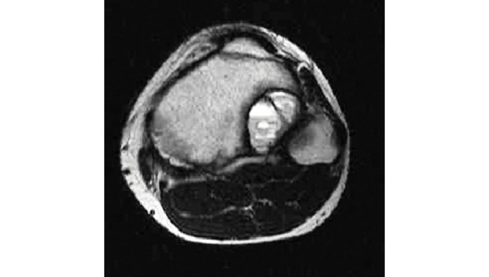
This is an axial T2 image, which shows a lesion in the posterolateral tibia. These appearances would be in keeping with an aneurysmal bone cyst.
That’s right. What’s the normal management for these?
First, it’s important to confirm the diagnosis and I would always discuss bony lesions of this type with a bone tumour MDT. In general, treatment of ABCs is with curettage and gratiing , but the recurrence rate can be as high as 50%. I am aware of newer therapies with promising results reported with the use of Denosumab, a RANKL inhibitor; however, its use in ABCs is currently off-label.
Figure 24.7 Aneurysmal bone cyst.
Other points
ABCs and GCTs may have similar appearances on plain X-rays and may only be reliably differentiated radiologically with MRI.
Traditionally , surgery was the mainstay of treatment.
‘Denosumab’ is a human monoclonal antibody that directly inhibits receptor activator of nuclear kappa B ligand (RANKL) signalling.
RANKL expression is seen in a variety of benign and malignant bone neoplasms and has higher than normal levels of expression in GCT and ABCs [10].
Among other things, ‘Denosumab’ is currently licensed for use in the treatment of skeletally mature adolescents and adults with GCT of bone [11].
improved pain, – in the case of spinal lesions – improvement in neurological symptoms [12].
Structured oral examination question 8#
Ewing’s sarcoma [5]
This is a histology slide taken from a biopsy of a tumour in the femoral diaphysis of a 16-year- old boy. What does this slide show (Figure 24.8)?
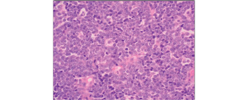
I’m no expert at histology, but I would describe the cells’ appearance as small, round and blue, and given the brief history you provided I suspect this may represent a Ewing’s sarcoma.
Excellent. What other features might this patient have presented with?
Patients usually present with pain and swelling related to the tumour. They usually present around the knee, with 25% occurring in the distal femur. Frequently, erythema, systemic pyrexia, a leukocytosis and a raised ES Rare also presenting features, which can incorrectly lead the unwary to a diagnosis of infection.
And what would be the characteristic features you’d look for on an X-ray?
Ewing’s sarcoma leads to a lytic, moth-ea ten appearance to the bone.
How would you investigate this further?
ACT chest is required to look for lung metastases, but, in Ewing’s sarcoma, a bone scan and bone marrow biopsy are also required to look for widespread bony metastases. Distant bone marrow involvement carries a significantly poorer prognosis.
In general terms, what is the management forEwing’s sarcoma?
There is a national video conference MDT for all cases of Ewing’s sarcoma, which recommends on management. Occasionally lesions are treated solely with chemo- and radiotherapy, usually in surgically inaccessible lesions around the pelvis. Five-year survival is 75% with a good response, but only 20% with a poor prognosis.
Figure 24.8 Ewing’s sarcoma.
Structured oral examination question 9#
Lipoma
This is an MR Iof apa tien t who has presented with a painless mass on the lateral aspect of his right elbow. To orientate you the round structure (labelled A) is the radial head. Tell me about the lesion adjacent to it, which is labelled B (Figure 24.9).
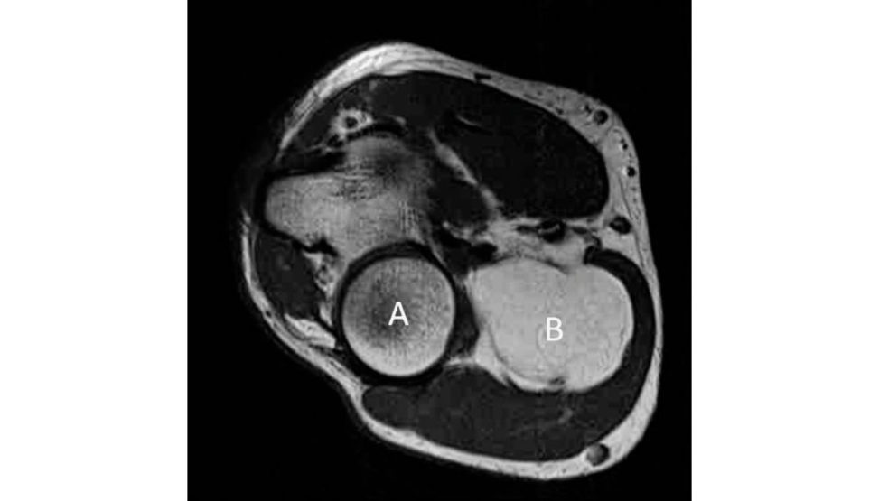
There is an intramuscular mass in the extensor compartment adjacent, and lateral, to the radial head.
What is a lipoma?
A lipoma is a benign tumour of mature adipocytes, identical to the surrounding adipose tissue, and showing liti le variation of cell size or shape.
And how would you treat this lesion?
Bland, innocent- looking lesions are usually treated with excision biopsy with a marginal margin. If there is any doubt, then a biopsy should betaken prior to excision. Histology of lesions below the fascia, like this one, often come back labelled as atypical lipomas by the histologist, despite very bland appearance on MRI.
What do you mean by an atypical lipoma?
The histology of such lesions shows variation of adipocyte size, in contrast to the bland adipocytes of a simple lipoma, and nuclear atypia, as well as the presence of lipoblasts. These lesions are benign and management is still with marginal excision, but they do have a low rate of local recurrence [8].
Figure 24.9 Lipoma.
Other points
Atypical lipomas (ALT) and well-differentia ted liposarcomas (WD Lare equivalent terms, describing lesions that are identical histologically.
Site-specific variations in behaviour relate only to surgical resectability.
They have no risk of metastasis – for these tumours the designation of AL T is preferred, as they do not behave like sarcomas.
In tumours that are large, pelvis, etc.), the risk of local recurrence, death are increased.
These lesions are best regarded as true sarcomas and the terminology of WD Lis more appropriate.
MDM2 amplification using fluorescent in-situ hybridization (FISH) is now a well-recognized method of differentiating WD Land AL T histologically.
Bibliography/Further reading
1. Welker JA, Henshaw RM, Jelinek J, Shmookler BM, Malawer MM. 2000;89(12):2677–2686.
2. Enneking WF, Spanier SS, Malawer MM. Cancer. 1981;47(5):1005–1022.
3. Enneking WF, Spanier SS, Goodman MA. J Bone Joint Surg Am. 1980;62(6):1027–1030.
4. NCCN. National Comprehensive Cancer Network Clinical Practice Guidelines in Oncology: So Tissueḁ
Sarcoma. V.2.2008. National Comprehensive Cancer Network. 2008.
5. Bullough PG. Orthopaedic Pathology. Fourth Edition . Edinburgh: Mosby; 2007.
6. Beckingsale TB, Gerrand CH. Osteosarcoma. Orthop Trauma. 2010;24(5):321–331.
Osteosarcoma: the addition of mur amyl tripeptide to chemotherapy improves overall survival – a report from the Children’s Oncology Group.
2008;26:633–638.
8. Beckingsale TB, Gerrand CH. The management of soft -tissue sarcomas. Orthop Trauma.
2009;23(4):240–247.
9. Jeys LM, Thorne CJ, Parry M, Gaston CL, Sumanthi VP, Grimer JR. Clin Orthop Relat Res.
2017;475(3):842–850.
10. Yamagishi T, Kawashima HOg ose A, et al. PL oSONE. 2016;11(5):e0154680.
11. Van der Hejden L, Dijkstra PDS, Blay JY, Gelderblom H.
12. Dubory A, Missenard G, Domont J, Court C. About nine cases. Spine (Phil aPa 1976).
2016;41(11):E654–660.