Chapter 27 Genetics and cell biology
Introduction#
The average orthopaedic trainee about to sit the.
This doesn’t need to be encyclopaedic, but candidates will need to have a sound grasp of disease inheritance and genetic disorders.
By comparison, mention genetic viva questions to any examiner and you get a slightly puzzled look back.
However, the subject does intermift ently appear in the vivas and therefore it is definitely.
Some genetic material will find its way into Section 1 SBA.
General information#
Nucleic acid structure (Figure 27.1)
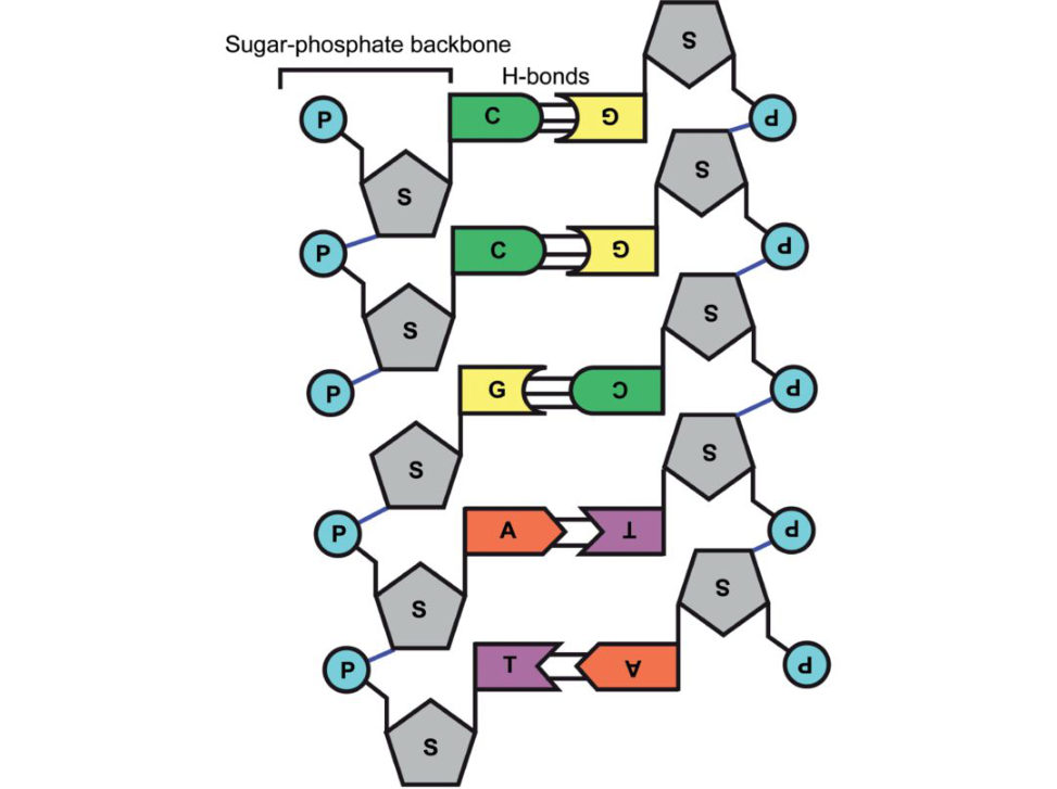
Figure 27.1 Nucleic acid structure.
There are two main types of nucleic acid, DNA (deoxyribonucleic acid) and RNA (ribonucleic acid), which each consist.
In DNA, there are two purine bases, adenine (A) and guanine (G)
(T) and cytosine (C) (Figure 27.2).
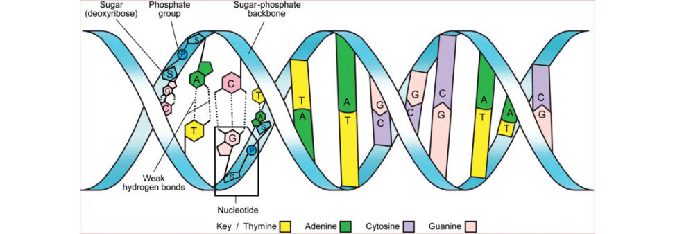
Figure 27.2 DNA structure.
RNA also contains adenine (A), guanine (G) and cytosine.
In DNA the sugar is deoxyribose, whereas in RN Aitis ribose. The nitrogenous bases are attached to.
Each unit of purine or pyrimidine base together.
A molecule of DN Ais composed of two nucleotide chains that are coiled clockwise 10 27.2).
The two chains run in opposite directions (i.e. 5ʹ to 3ʹ for one and 3ʹ to 5ʹ for the other) and.
Pedigree charts
Pedigree charts can be complicated to.
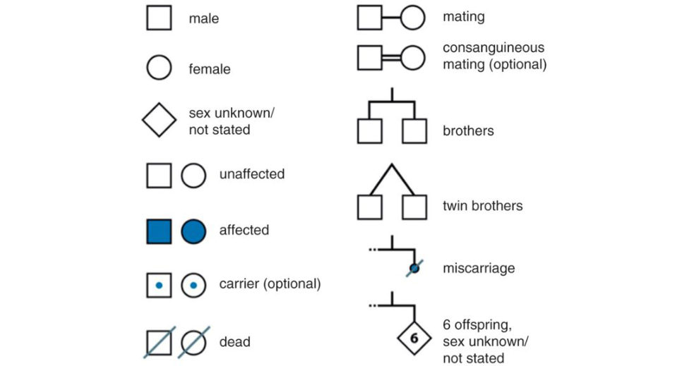
A standard set of symbols are used (Figure 27.14). Roman numerals are used for each generation, starting with the earliest, and Arabic numerals are used to indicate each individual within a generation (numbering from the left).
Single gene disorders are due to mutations in one or both copies or alleles of an autosomal gene or to.
Structured oral examination question 1#
Autosomal dominant (AD) inheritance
This is much easier to explain if candidates are able to 27.3).
What pattern of inheritance does the pedigree chart show and why (Figure 27.4)?
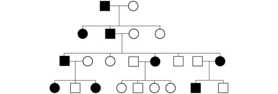
This is autosomal dominant because: At conception, each child has a 1 in 2 (50%) chance of inheriting the condition. It is equally likely that a child will receive the mutant or normal allele from the affected parent. On average there is a 1 in 2 or 50% chance that each child of a heterozygous parent will inherit the gene mutation. There is usually a variation in time of onset and severity of condition with AD traits. This is likely to be due in part to the effects of other ‘modifier’ genes and also lifestyle/environmental factors.
Choose an autosomal dominant condition and discuss the genetics.
A gift if you have done your homework. The three most obvious disorders to choose are (1) achondroplasia, (2) osteogenesis imperfect aor (3) neurofibromatosis. 1 Candidates are throwing away easy scoring opportunities. The other situation is that the clinical condition to discuss has already been decided beforehand.
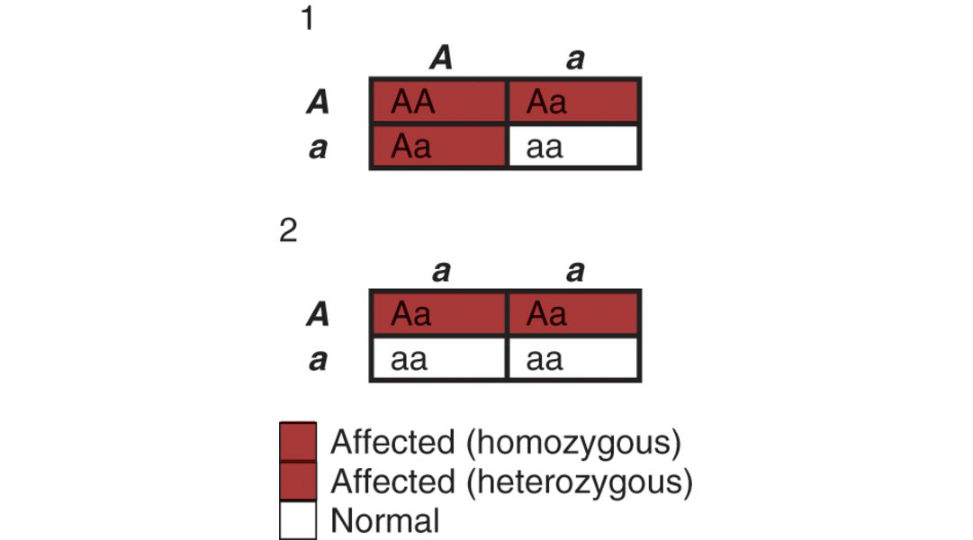
Figure 27.3 Punneft square demonstrating inheritance of an autosomal dominant trait: (1) two heterozygous parents, (2) one heterozygous and one unaffected parent.
Figure 27.4 Pedigree chart of AD inheritance, one affected heterozygous and one unaffected parent. The disease is passed from father to son – this almost never happens with X-linked traits. The disease occurs in three consecutive generations – this almost never happens with recessive traits.
Achondroplasia
About 80% of people with achondroplasia have the condition as the result of a new mutation;
Each child of someone who has achondroplasia has a 1 in 2 (50%) chance of inheriting the condition.
If both parents have achondroplasia, and a child inherits a copy of the.
The gene for achondroplasia is on the 4.
The condition is the result of a mutation
3 (FGFR3), which is a key component of.
The main defect isabnormal endochondral bone.
Clinical features of achondroplasia may be discussed in more detail and these can easily be mugged.
What is the genetic mutation responsible for this condition, and what effect does this have?
Achondroplasia is caused by a single point mutation in the gene encoding the fibroblast growth factor receptor 3 (FGFR3). T wo mutations in the FGFR3 gene are responsible for 99% of cases of achondroplasia. Soluble FGFR3 has successfully been used in mice to act as a decoy for FG Fin order to restore normal growth in achondroplasia.
What is the mode of inheritance of this condition?
It is autosomal dominant.
This child’s parents do not have this condition. How is this possible?
A large proportion (80%) of cases occur due to a spontaneous (new) mutation.
Osteogenesis imperfecta
There is a qualitative or quantitative defect of type 1 collagen synthesis. Originally classified by.
The vast majority of cases are autosomal dominant.
Type 1 collagen is the major extracellular protein.
The structural unit of type 1 collagen is called tropocollagen and is a heterotrimer composed of three polypeptide chains. Two chains are pro-α1(I) and the third chain is pro-α2(I).
The triple helical structure is not the same as the pro-α helix that is.
The collagen triple helix forms because both the α1 and α2 chains contain repeat sequences of amino acids (–Gly–X–Y), where Gl yis glycine, X is proline andY is usually hydroxyproline.
A missense mutation 4 leading to the replacement of even one Gl yin the repeating (Gly–X–Y)n sequence by a larger residue may lead to a pathological condition.
The pro-α1(I) chain is encoded from the COL1A1 gene on chromosome 17 pro-α2(I).
Mutations in the COL1A1 gene on chromosome 17 COL1A2.
In the type 1 condition the COL1A1 mutant pro-α1(I)
There is only 50% production of normal pr o-α1(I) chain.
In more severe forms of the condition there are mutations of either the COL1A1 COL1A2.
Structured oral examination question 2#
Autosomal recessive (AR) inheritance
Can you please draw a Punneft square demonstrating an autosomal recessive trait (Figure 27.5).
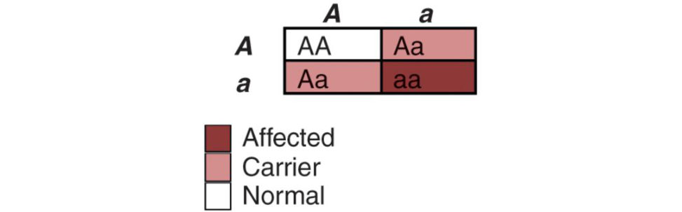
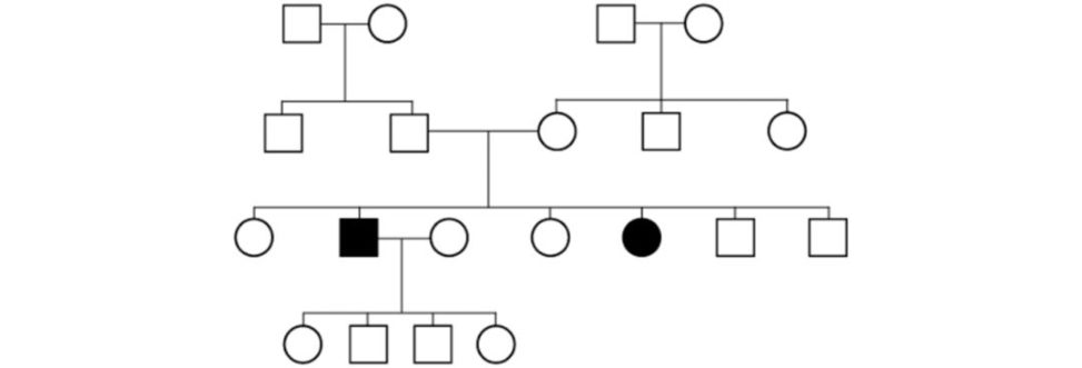
Recessive means two copies of the gene are necessary to have the trait/disease. If the parents are both carriers the risk of them having an affected child is 25% (1/4) and the risk of them having a child who is a carrier is 50%. Unaffected adult offspring of carrier parents have a 2/3 risk of carrier status. Alternatively candidates may be shown a pedigree chart of autosomal recessive inheritance and asked to identify the pattern of inheritance (Figure 27.6). On average, the recurrence risk to the unborn sibling of an affected individual is 1/4. Within the completely unaffected siblings of an affected individual the probability of being a carrier is 2/3.
Can you describe an autosomal recessive disease?
Obvious choices include (1) sickle cell anaemia, (2) mucopolysaccharidoses – all except Type.
Figure 27.5 Punneft square demonstrating autosomal recessive inheritance.
Figure 27.6 Pedigree chart of autosomal recessive inheritance.
Sickle cell disease
Sickle cell disease describes a group of disorders caused by a mutation in the beta globin gene (HBB). SC Ais the commonest of these diseases and is caused by homozygous point mutations in the HBB gene on the short arm of chromosome 11.
Heterozygous carriers of HBB mutations (‘sickle cell trait’) have a selective advantage (heterozygote advantage), due to their resistance to malaria. Hence heterozygotes have increased chances of survival in malaria-prevalent areas.
at high altitude) or dehydrate, an important factor.
Clinical features
SC Ais characterized by episodes of pain owing to vaso-occlusive events,.
Any organ may be affected but most commonly bones (ON/osteomyelitis).
Management includes analgesia, fluids, oxygen and in severe cases exchange transfusion.
Structured oral examination question 3#
X-linked inheritance
Can you draw the Punneft square for an X-linked dominant condition ( Figure 27.7 and 27.8)?
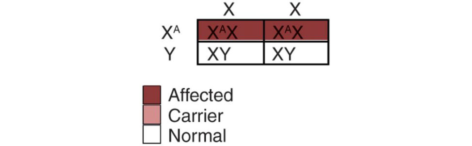
[While drawing the Punneft square.] If the father is affected his sons will be unaffected and all his daughters will be affected (Figure 27.7).
What is the mode of inheritance here (Figure 27.9)?
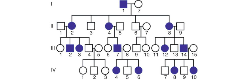
Figure 27.7 Punneft square showing inheritance of an X-linked dominant trait (normal mother and affected father).
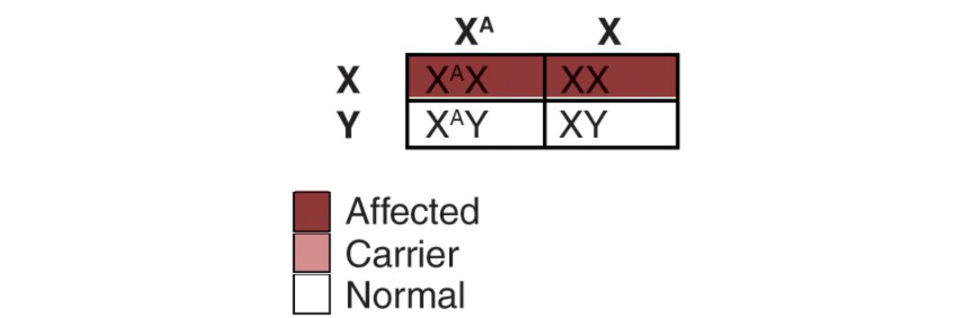
Figure 27.8 Punneft square showing inheritance of an X-linked dominant trait (affected mother and normal father).
Figure 27.9 X-linked dominant inheritance. The key for determining if a dominant trait is X-linked or autosomal is to look at the offspring of the mating of an affected male and a normal female. If the affected male has an affected son, then the disease is not X-linked. All of his daughters must be affected if the disease is X-linked dominant.
This is X-linked dominant inheritance. Hallmarks of X-linked dominant inheritance include: Matings of affected females and normal males result in 1/2 of the sons being affected and 1/2 of the daughters being affected. In the general population females are more likely to be affected than males, even if the disease is not lethal in males.
What is the mode of inheritance here (Figure 27.10)?
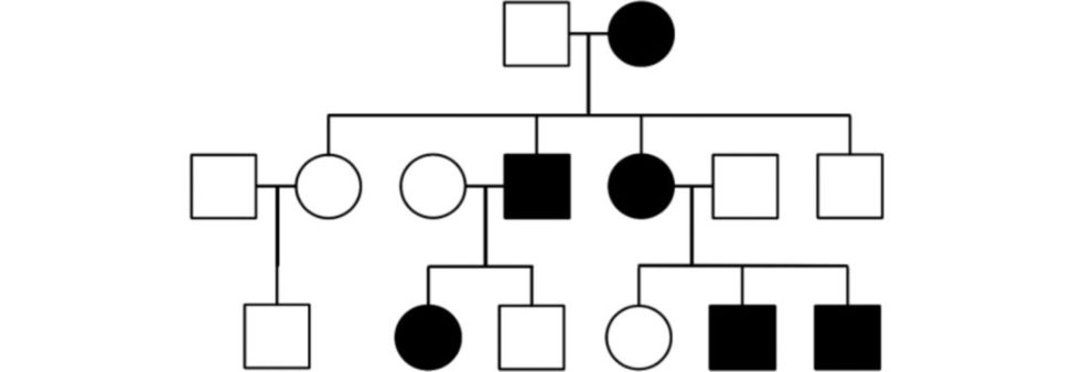
This is the opposite situation to Figure 27.9 where the male parentis affected. This is still X -linked dominant inheritance. An X-linked dominant trait does not skip generations. Affected mothers (if heterozygous) will pass the trait on to half of their sons and half of their daughters.
Do you know of an example of an X-linked dominant inheritance condition?
Hypophosphataemic rickets (vitamin D-resistant rickets).5 The condition is caused by mutations in the PHEX geneP HEX.
In addition, the absence of PHEX enzymatic activity may cause accumulation ofos teopontin (a.
Figure 27.10 X-linked dominant inheritance. Parent female is affected. Affected father does not pass disease on to son.
Clinical features
Childhood rickets with growth retardation and poor dental development.
In middle age, mineralization of spinal ligaments and thickening of neural arches. Loss of mobility
D.
The condition is caused by mutations in the PHEX gene. The change created in the gene is a loss-of-function mutation, resulting in reduced breakdown and circulatory clearance of FGF23. FGF23 acts on the kidney to cause increased phosphate excretion and decreased alpha-1 hydroxylase activity . Conradi–Hunermann chondrodysplasia punctata (due to a mutation in the gene encoding EBP).
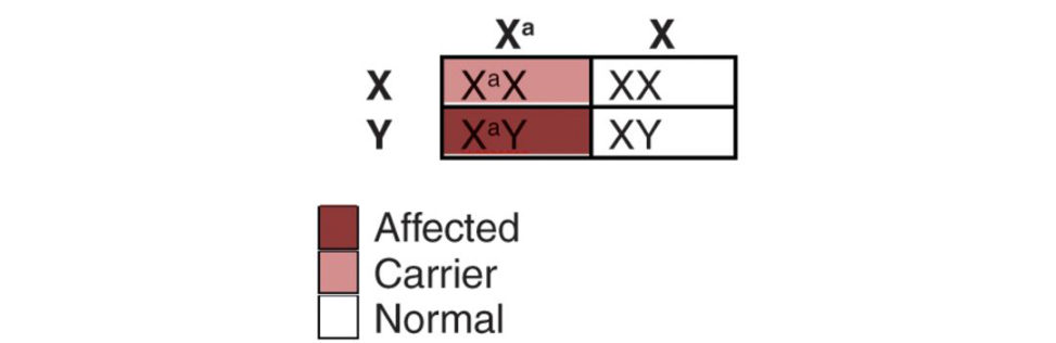
Can you draw the Punneft square for an X-linked recessive condition?
Figure 27.11 and 27.12.
What is the mode of inheritance here (Figure 27.13)?
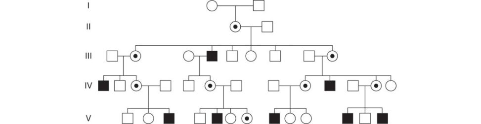
This is X-linked recessive. As with any X-linked trait, the disease is never passed from father to son. For a carrier female, with each pregnancy there is a one in two (50%) chance her sons will inherit the disease allele and a one in two (50%) chance her daughters will be carriers. Affected males transmit the disease allele to all of their daughters who are then carriers, but to none of their sons.
Figure 27.11 Punneft square showing inheritance of an X-linked dominant trait (affected mother and normal father).
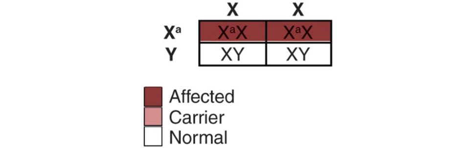
Figure 27.12 Punneft square showing inheritance of an X-linked dominant trait (normal mother and affected father).
Figure 27.13 X-linked recessive inheritance.
Duchenne muscular dystrophy (DMD)
Incidence approximately 1 in 4000 boys.
This is one of the dystrophinopathies caused by a mutation in the dystrophin gene (Xp21). The dystrophin protein provides structural stability to the dystroglycan complex of the muscle cell membrane, and its function is lost as a result of the mutation.
Clinical features
Age of onset is usually before 6 years. Progressive.
Compensatory toe walking is.
Frequent falls/fatigue.
Speech delay and difficulty with motor skills.
Lumbar lordosis/scoliosis.
Usually wheelchair-bound by 12 years 25.
Gower’s sign positive the child is unable to jump up quickly from a.
Other examples of X-linked recessive inheritance:
Becker muscular dystrophy.
Mucopolysaccharidosis Type II (Hunter’s syndrome).
Haemophilia A. Genetic defect in factor VIII.
SED tarda.
Pick one family pedigree and tell me the mode of inheritance (Figure 27.15).
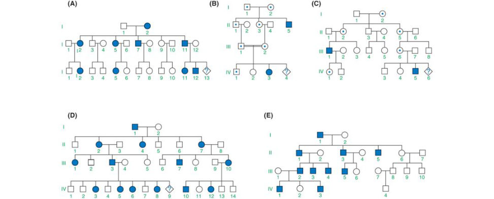
Be able to justify your answer.
Figure 27.14 Symbols for constructing a family tree.
Figure 27.15 Family tree for various modes of inheritance. A, autosomal dominant; B, autosomal recessive; C, X-linked recessive; D, X-linked dominant; E, Y-linked inheritance.
Structured oral examination question 4#
Stem cells
Potentially an awkward C-list topic, especially if a.
What are stem cells?
Stem cells represent unspecialized cells that have the ability to.
What two properties must a stem cell demonstrate?6
If stem cells could not self-renew, tissues would runout of replacement cells for those that had died. This requires stem cells to be either totipotent or pluripotent – to be able to give rise to any mature cell type.
What are the different types of stem cells that you know?
The two main types of stem cells are: 1. 2. Induced pluripotent stem cells (IPSCs). Nuclear transplant stem cells (ovasomes). Parthenote stem cells.
What do you mean by pluripotent?
Pluripotent cells have the capacity to differentiate into any cell type in the body. Multipotent cells can develop into more than one cell type but are more limited incapacity than pluripotent cells. They can form many types of cell in a given lineage, but not cells of other lineages.
Can you think of any uses of stem cells in orthopaedics?
Stem cells have become a focus of regenerative medicine. The goal of stem cell therapy is to replace or replenish diseased tissue through the localized differentiation of transplanted stem cells into cells which advance the healing processor directly restore the tissue physically. 1. Stem cells assist with growth factor release and alteration of the anatomic microenvironment to facilitate regeneration and repair of the chondral surface. Due to their role in inhibiting the catabolic activity of matrix metalloproteinases (MMP), mesenchymal stem cells (MSCs) have been shown to have a beneficial effect in OA. 2. 3. 4. 5. Spinal cord injury7 There has been recent research into cell-based therapies for spinal cord injury. 6. Meniscal injury Isolated case reports exist of meniscal regeneration after percutaneous injection of autologous ASCs into an adult human knee.8 It is not clear whether this is a direct action of the mesenchymal-based cells or is rather mediated by secretion of certain stimulating factors on the existing meniscal tissue.
Stem cells have also been added to modify the biomechanical environment.
7. Intervertebral disc disease
Various clinical trials have been.
Percutaneous stem cell mediated disc regeneration has the potential to establish itself as.
8. Spinal fusion
Pseudoarthrosis remains a pressing issue occurring in 13–41.4% of patients undergoing spinal fusion.
MSCs and adipose tissue derived stem cells (ADSCs) have both demonstrated a significant positive effect on spinal fusion in a number of experimental models.
9. Physeal injury/defects
Several animal models have investigated the use of stem cells combined.
10. Osteonecrosis
Core decompression and injection of isolated stem cells have been used in early stages of ON hip.
What are your concerns with the use of stem cells in orthopaedics?
MSCs have been reported to promote tumour growth and metastases. There is very limited clinical experience with pluripotent stem cells (embryonal stem cells and IPSC). Retroviruses may be used to generate human IPSCs. These viruses are genetically altered to express the genes that are required for transformation in to an IPSC. Applying this genetic reprogramming, the used viruses can integrate into the cell genome.
the cells may contain multiple viral integration sites in their genomes.
Cost.
Microbial contamination during cell amplification.
Controlling stem cell differentiation.
Control of their proliferation and differentiation in to.
Lack of adequate vehicles/scaffolds.
Integration with local tissues.
Immunological rejection and disease transmission (if allogeneic).
Potential modulation of host.
Continuous cell amplification of.
Notes
1. This will usually end upmaking the viva.
2. Ward JG argan A, Smiths onS, Atherton G. 2013;27(4):229–232.
3. an expanding panorama of variants. 1981;159:11–25.
4. A missense mutation is a point mutation in which a single.
5. Best to stick to hypophosphatemic rickets as the rest.
6. Much better if a candidate volunteers this.
7. Schroeder GD, Kepler CK, Vaccaro AR. 2016;24(4):266–275.
8. Pak J, Lee JH, Lee SH. Regenerative repair of damaged.