Chapter 31 Drawings for the FRCS (Tr & Orth)
James Widnall, Catherine McCauley and Lyndon Mason
Drawing has always been an integral part of the FRCS examination, be it as a method to understand a complex process during revision or as a tool for explanation in the viva itself.
having recently been through the examination ort aught on this subject for many years.
Some of the drawings are deliberately schematic in order to simplify the subject matter and reinforce key points.
We hope, though, that by using these drawings to either act as an aide memoir during revision, or indeed to explain difficult concepts.
Good luck.
Tips on drawing#
An exam is not just about knowledge but the presentation of that knowledge. There is a lot of paper at every station, and sometimes it’s useful to draw even if not asked to do so. Remember the old adage ‘a picture is worth a 1000 words’.
whether as an aid to explain an answer or as the result of being asked to do so.
1. Drawing small does not show confidence.
2. Work from the outside in. Simplify to the bare image. For example, if drawing a cross-section of a limb,.
3. Draw schematic . The examiners will not.
4. Speak while you draw. You have limited timet o earn.
Anatomy#
Anatomy is assumed knowledge for surgeons; it is, after all, a subject matter we deal with everyday. Thus, an anatomy question in the exam should be dealt with confidently and efficiently, otherwise.
NB: some of the illustrations below are of cross-sectional anatomy in the limbs. Not only do these convey a knowledge of surrounding structures and.
We have given you both anatomically correct and schematic diagrams for these.
Upper limb
Brachial plexus (Figure 31.1)
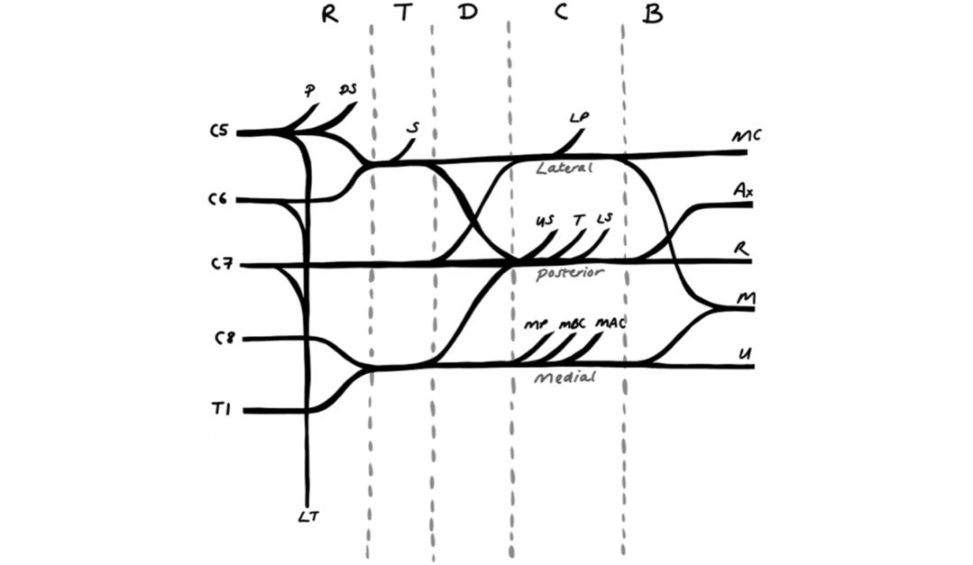
Figure 31.1 R, roots; T, trunks; D, divisions; C, cords; B, branches. P, phrenic nerve (contribution); DS, dorsal scapularS, suprascapular; LP, lateral pectoral; MC, musculocutaneous; US, upper subscapular; T, thoracodorsal; LS, lower subscapular; AX, axillary; R, radial; MP, medial pectoral; MBC, medial brachial cutaneous (of arm); MAC, medial antebrachial cutaneous (of forearm); U, ulnar; 1st IC: first intercostal; LT, long thoracic nerve (of Bell).
The candidate not only needs to know the anatomy of the brachial plexus but also how to apply it to a clinical picture/examination. There are a number of
Youtube videos that teach.
The candidate can also demonstrate whether
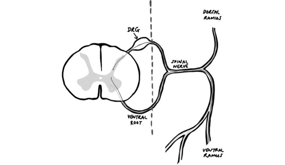
(post-ganglionic) the dorsal root ganglion (see Figure 31.2).
Dorsal root ganglion (Figure 31.2)
Figure 31.2 DRG, dorsal root ganglion.
Figure 31.2 clearly shows where the division between pre- and post-ganglionic injuries exists. Pre-ganglionic injuries may present with Horner’s syndrome,.
Erb’s point (Figure 31.3)
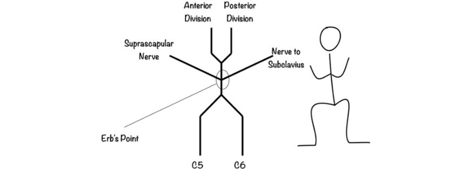
Figure 31.3 Erb’s point.
Not to be confused with the cardiology Erb’s point (third intercostal space on the left sternal border where S2 heart sound is best auscultated)
Erb’s point that concerns us is a site at the upper trunk of the brachial plexus located 2–3 cm above the clavicle. Erb’s point is formed by the union of the C5 and C6 nerveroot sAt the nerve trunk, branches of suprascapular nerve and the nerve to the subclavius also merge.
Humeral spaces (Figure 31.4)
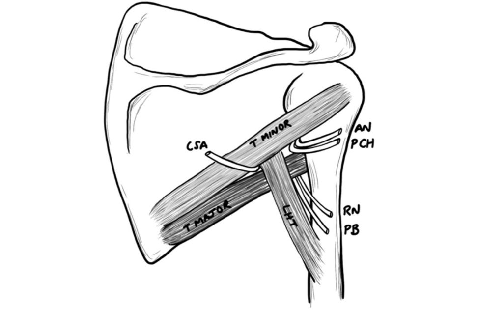
Figure 31.4 Humeral spaces.
A useful aide memoir for this is to place the index and middle fingers of your right hand at right angles to your left hand index and middle fingers to create the same shape.
Clinically, these spaces are important when performing the posterior approach to the shoulder, knowing that finding the interval between infraspinatus.
Humerus cross-section (Figure 31.5)
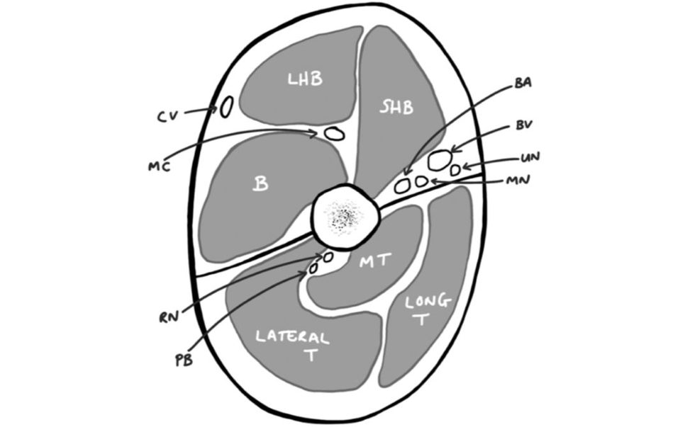
Figure 31.5 CV, cephalic vein; MC, musculocutaneous nerve; BA, brachial artery; BV, basilica vein; UN, ulnar nerve; MN, median nerve; RN, radial nerve; PB, profunda brachii.
LHB, longhead biceps; SHB, shorthead biceps; B, brachialis.
Lateral T, lateral head of triceps; MT, medial.
The above illustration can be used.
1. Anterior – retract biceps laterally, identify radial.
2. Posterior – develop interval between long and lateral heads of triceps,.
Schematic drawing (Figure 31.6)
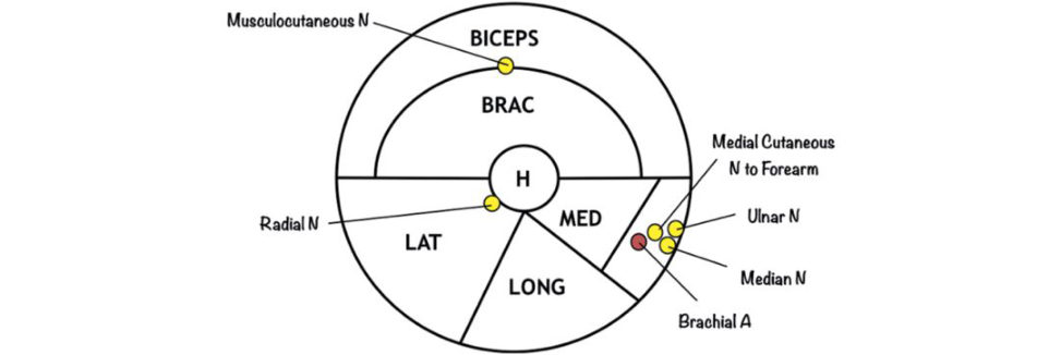
1. First draw a large circle and label.
2. Add the humerus centrally and divide the.
It initially looks like a Pokemon ball. By drawing the mid-humeral cross-section you.
3. The musculocutaneous nerve lies in the plane between these muscles and supplies the medial half of the muscle.
4. In the posterior compartment the three heads of.
5. The radial nerve runs in the spiral.
The ulnar nerve, median nerve and medial cutaneous nerve of the.
Figure 31.6 Schematic drawing of humerus cross-section.
Mid-forearm cross-section (Figure 31.7)
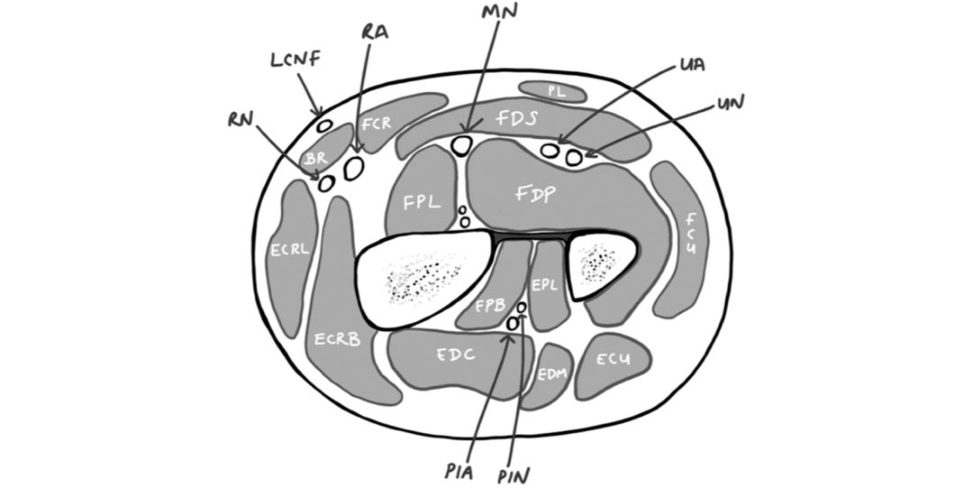
Figure 31.7 RN, radial nerve; LCNF, lateral cutaneous nerve of the forearm; RA, radial artery; MN, median nerve; UA, ulnar artery; UN, ulnar nerve; PIA, posterior interosseous artery; PIN, posterior interosseous nerve.
BR, brachioradialis; FDS, flexor digitorum superficialis; FDP, flexor digitorum profundus; FCU, flexor carpi ulnaris.
ECRL, extensor carpi radialis longus; ECRB, extensor carpi radialis brevis; EDC, extensor digitorum communis;
Schematic drawing (Figure 31.8)
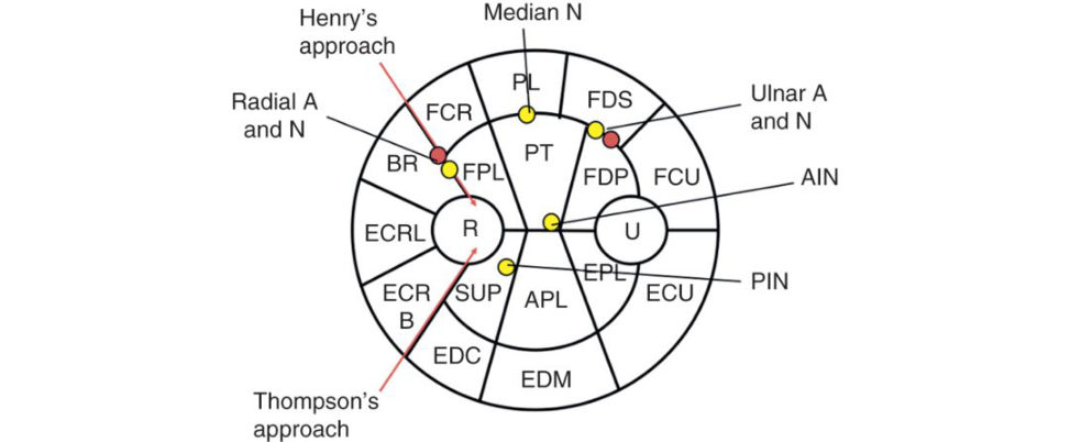
Figure 31.8 Schematic drawing of mid-forearm cross-section.
Drawing the forearm cross-section (Figure 31.8) is a daunting.
1. Again, draw a large circle.
2. Draw the radius and ulna and divide the forearm.
3. Divide the compartments into superficial and deep and then divide the anterior superficial into four, deep into three;
4. BR lies anterior-most, followed byE CRL and ECRB.
5. Thinking of Henry’s approach, FCR can then be added.
The other superficial muscles.
6. We know the SRN and radial artery.
7. As half of the FDP is supplied by the ulnar nerve, this is where the ulnar nerve and artery lie.
8. The remainder of the deep compartment depends on the level, so considering the.
9. The median nerve lies deep to palmaris longus and.
10. On the other side of the superficial compartment lies EDC, which forms the interval for Thompson’s posterior approach to the radius.
11. The remaining extensors depend on the level.
12. In the deep layer, the supinator lies around the radius; it is likely to only be partially present.
13. The PIN lies within the supinator.
14. Finally, the common approaches to the forearm
Thompson’s between ECRB and EDC.
Approaches to the midshaft radius (Figure 31.9)
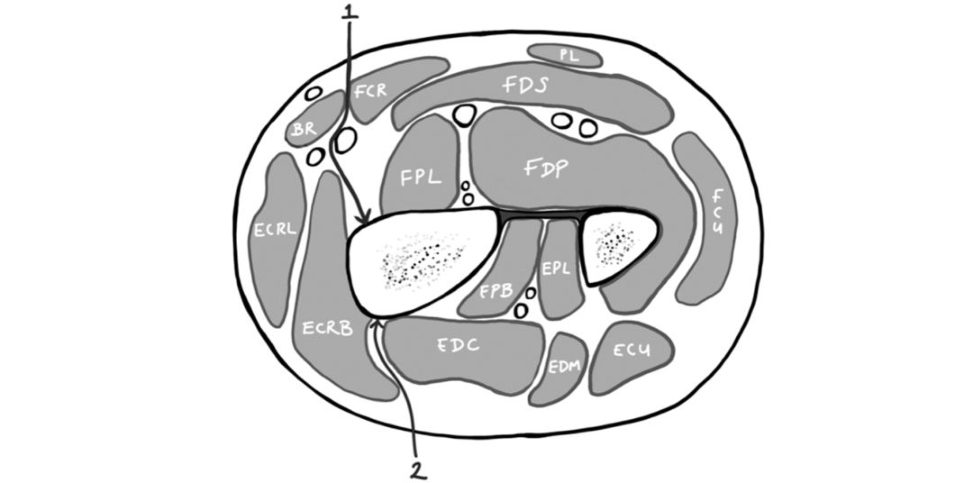
Figure 31.9 Approaches to the midshaft radius.
1. The modified Henry’s approach (which we use for distal radius fractures) differs bygoing through the bed of the FCR, creating a plane between the FC Rand radial artery.
2. Thompson’s approach – develop the interval between.
Radius muscle insertions
Radius muscle insertions are often combined with the cross-section of the forearm. There is one supinator (excluding the biceps attachment), two pronators, and two interspersing long flexors to the digits. The supinator for the ears, the flexors (FP Land FDS) for the body and the pronators (PQ and PT) for the legs (Figure 31.10).
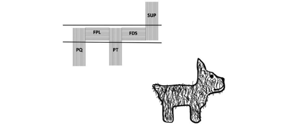
Figure 31.10 Radius muscle insertions.
The deep dissection of Henry’s approach to the radius requires the supinator to be incised at its insertion on the radius, when the forearm is in full.
Pronator teres in the middle third of the radius.
Hand and wrist
Carpal tunnel
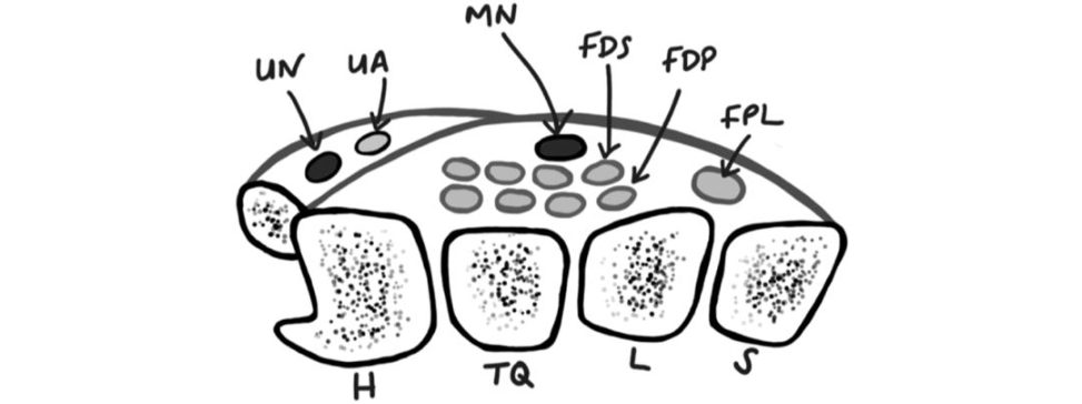
Figure 31.11 UN, ulnar nerve; UA, ulnar artery; MN, median nerve; FDS, flexor digitorum superficialis; FDP, flexor digitorum profundus; FPL, flexor pollicis longus.
H, hamate; Tq, triquetrum; L, lunateS scaphoid.
The contents of the carpal tunnel are easily tested. This diagram (Figure 31.11) can be a quick way to detail the necessary knowledge.
The carpal tunnel spans from the scaphoid tubercle and trapezium ridge to the hook of hamate and pisiform. The roof is the transverse carpal.
As well as the cross-sectional anatomy the candidate should be familiar with the branches of the median nerve, e.g.
extraligamentous 75% of time, transligamentous 12%).
Extensor compartments (Figure 31.12)
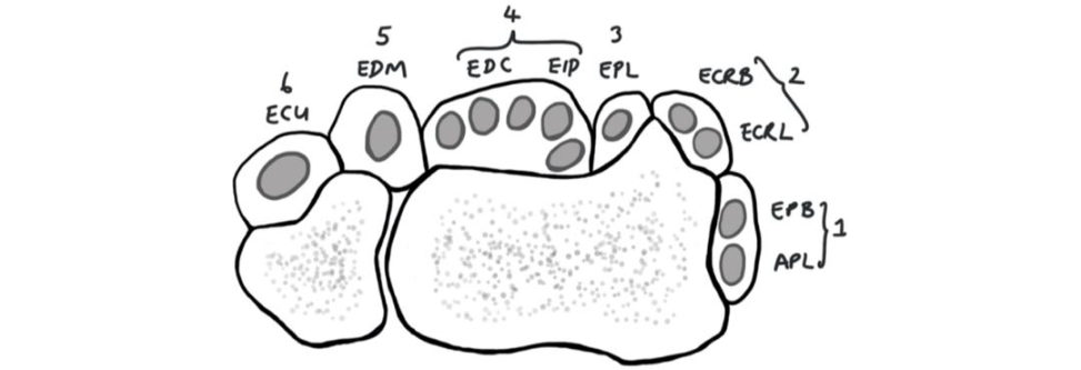
Figure 31.12 Compartment 1: APL, abductor pollicis longus; EPB, extensor pollicis brevis.
Compartment 2: ECRB, extensor carpi radialis brevis.
Compartment 3: EPL, extensor pollicis longus.
Compartment 4: EIP, extensor indicis propius.
Compartment 5: EDM, extensor digift minimi.
Compartment 6: ECU, extensor carpi ulnaris.
It also details the proximity of EPL to Lister’s tubercle, which can contribute to atiritional rupture of the tendon.
Ulnar canal (Figure 31.13)
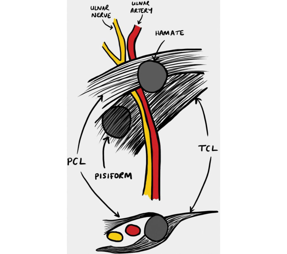
The roof of the canal is made up of the superficial palmar carpal ligament (PC Land floor by the transverse carpal ligament (TCL).
Anatomy is a possible question especially duet ozones.
Zone 1: proximal to bifurcation of nerve (mixed motor.
Zone 2: Surrounds deep motor branch (motor only,.
Zone 3: Surrounds superficial sensory branch (sensory only,.
Figure 31.13 Ulnar canal.
Sites of ulnar nerve compression (Figure 31.14)
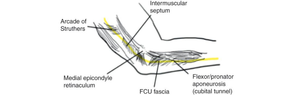
Management of ulnar nerve compression depends on an accurate diagnosis, yet localizing the site of nerve compression can be challenging. The accepted sites of potential ulnar nerve compression are depicted here: arcade of Struthers, the medial intermuscular septum, the bony retrocondylar.
Figure 31.14 Sites of ulnar nerve compression.
Finger cross-sectional anatomy
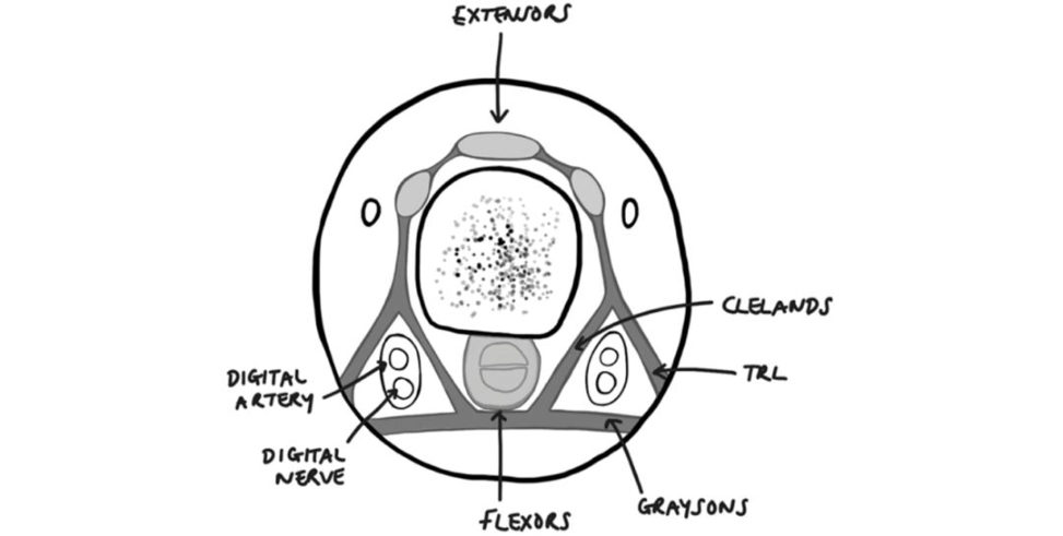
Figure 31.15 TRL, transverse retinacular ligament.
This axial section (Figure 31.15) shows the relationship of the neurovascular bundles to that of the fascial sheets in the finger.
Grayson’s ligament (remember Cleland’s ligament is not involved in Dupuytren’s disease) can.
Nail anatomy (Figure 31.16)
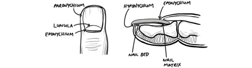
Figure 31.16 Nail anatomy.
With relative ease this viva topic could quickly proceed to high levels talking about potential incisions to extend wounds, nail bed repair or flap coverage.
Lower limb
Blood supply to neck of femur (Figure 31.17)
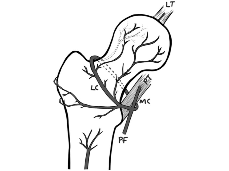
Figure 31.17 LT, ligamentum teres; PT, psoas tendon; PF, profunda femoris; MC, medial circumflex; LC, lateral circumflex.
The blood supply to the femoral head is a large part of what we base our.
The profunda femoris splits into medial and lateral circumflex arteries (so named in their relationship to the psoas tendon as shown). The anterior arises from the lateral circumflex.
A small branch from the obturator artery passes through the ligamentum teres.
Femoral triangle (Figure 31.18)
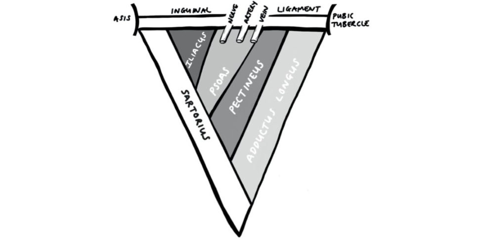
Figure 31.18 ASIS, anterior superior iliac spine.
The femoral triangle (Figure 31.18) is a common.
Boundaries: medial – medial border of adductor longus;
– inguinal ligament.
Roof: fascia lata.
Contents: femoral nerve, artery, vein, inguinal lymph nodes.
Floor: iliacus, psoas, pectineus, adductor longus.
Hip cross-section anatomy (Figure 31.19)
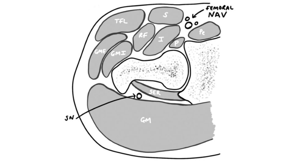
Using the retained knowledge from the femoral triangle, we already have the medial aspect of the cross-section of the hip. The only structure not represented is the adductor longus, but this is due to its origin being located below the hip, at the anterior inferior iliac spine.
Figure 31.19 TFL, tensor fascia lataS, sartorius; Pe, pectineus; P , psoas; I, iliacus; RF, rectus femoris; GMI, gluteus minimus; GME, gluteus medius; SER, short external rotators; GM, gluteus maximus; SN, sciatic nerve.
Approaches to the hip (Figure 31.20)
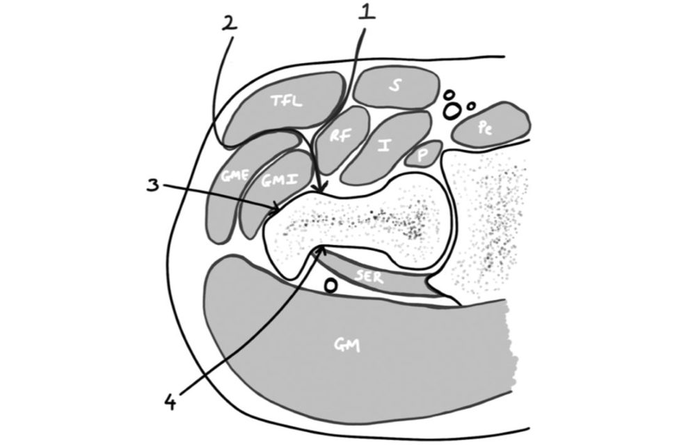
Figure 31.20 Approaches to the hip.
Again, this knowledge of cross-sectional anatomy can be applied to quickly.
1. Smith Peterson – superficial plane developed between the sartorius and tensor fascia lata, being.
Deeper dissection is between.
2. Watson-Jones – develops the plane between the.
3. Lateral approach – after incising the fascia lata the abductors (gluteus medius/minimus).
4. The short external rotators can then be elevated from the insertion to gain access to the posterior capsule, protecting thes cia tic nerve throughout.
Adductor/Hunter’s canal (Figure 31.21)
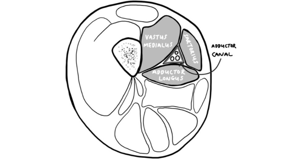
Figure 31.21 Adductor/Hunter’s canal.
The adductor canal, or Hunter’s (John Hunter, Scotish surgeon, 1728–1793) canal extends from the apex of.
Contents: superficial femoral artery, femoral vein, saphenous nerve.
Boundaries: medial wall – sartorius; posterior wall –.
Mid-thigh cross-section (Figure 31.22)
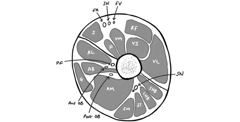
Figure 31.22 FA, femoral artery; SN, saphenous nerve; FV, femoral vein; SN, sciatic nerve; Post OB, posterior branch of obturator; Ant OB, anterior branch of obturator; PF, profunda femoris.
S, sartorius; VI, vastus intermedius; RF, rectus femoris.
AL, adductor longus; AB, adductor brevis; LHB, longhead biceps femoris; SHB, shorthead biceps femoris.
The most common approach to the thigh is that of direct lateral where the surgeon incises the fascia before either.
Schematic (Figure 31.23)
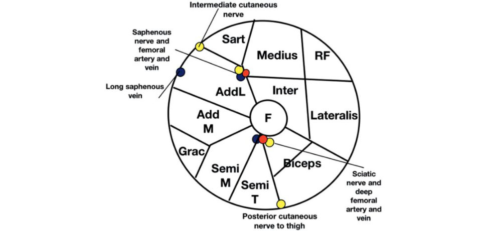
1. Draw a large circle and add the femur centrally.
2. On the peripheries either side of the adductor compartment, add the sartorius and gacillis muscles, which lie in their own muscle fascia.
3. Divide the adductor compartment into two and.
4. Add in the muscles, with the adductor compartment containing adductor longus and magnus, the anterior compartment containing the quadriceps.
5. Add NV structures as shown.
Figure 31.23 Schematic drawing of mid-thigh cross-section.
Lower leg cross-section (Figure 31.24)
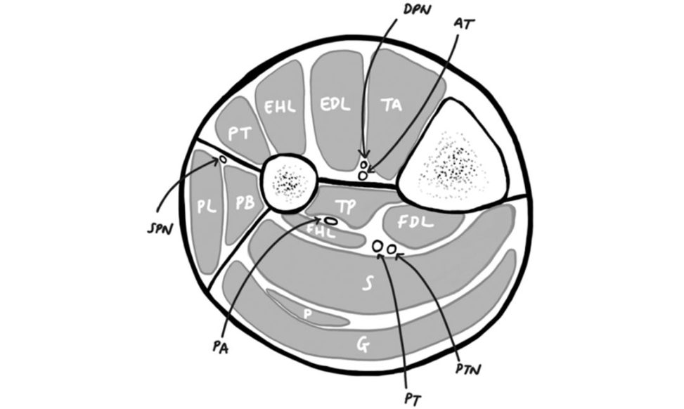
Figure 31.24 Anterior compartment: PT, peroneus tertius; EHL , extensor halluces longus; EDL, extensor digitorum longus; TA, tibialis anterior; DPN, deep peroneal nerve; AT, anterior tibial artery.
Posterior compartment (deep): FDL, flexor digitorum longus; PTN, posterior tibial nerve.
Posterior compartment superficialS, soleus;
Lateral compartment: SPN, superficial peroneal nerve.
It is important to note that while the peroneal artery runs in the deep posterior compartment, it actually supplies the lateral compartment via perforators.
The compartments of the lower limb are often asked in the context.
Schematic (Figure 31.25)
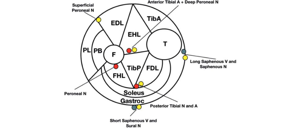
Figure 31.25 Schematic drawing of lower leg cross-section.
1. Draw a circle and add the tibia.
2. Add the intermuscular septum.
3. Divide and label the four compartments (anterior,.
4. Divide the anterior and posterior deep compartment into.
5. Add in the muscles; anterior compartment (tibialis anterior, extensor hallucis longus and extensor digitorum longus), deep posterior compartment (tibialis posterior, flexor digitorum
FHL), B close to bone) and posterior superficial (soleus and gastrocnemius).
6. Add in the anterior tibial nerve and artery (anterior to intraosseus membrane), posterior.
For extra marks, add in the short and.
Basic science#
Biological materials
Articular cartilage (Figure 31.26)
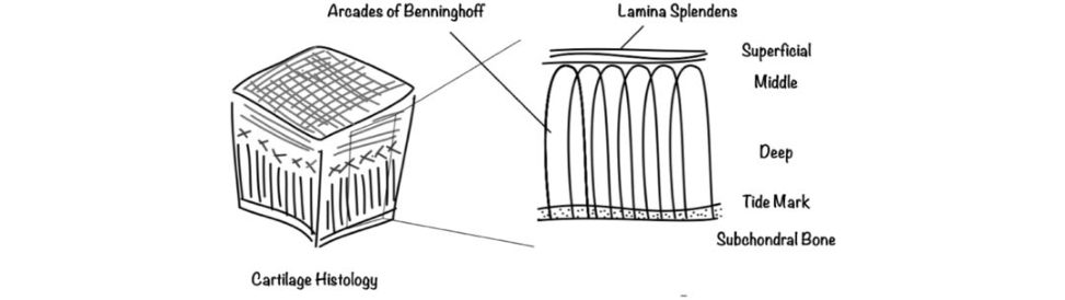
Figure 31.26 Articular cartilage.
Articular cartilage is a common viva question given that it is at.
There is an outer protective layer of the lamina splendens.
This covers the superficial layer where the collagen fibres are parallel to the articular surface in order to resist shear forces. Here, the proteoglycan concentration is the highest, with there being the fewest collagen fibres and water molecules. The calcified zone anchors the cartilage to bone via hydroxyapatite crystals. This zone is mostly type X collagen, unlike the other zones which are mainly type II.
Most trainees memorize the histological features with the zones representing columnar areas, cross-hatching and parallel fibres.
The candidate needs to know the difference between OA and.
Differences in ageing and osteoarthritis
This is best remembered if you imagine an elderly individual who is stiff and dry (Figure 31.27). This is the opposite for OA.
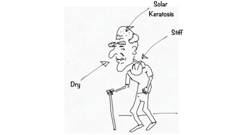
Figure 31.27 Differences in ageing and osteoarthritis.
Proteoglycans (Figure 31.28)
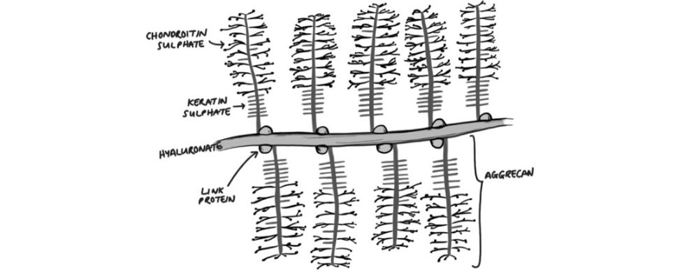
Figure 31.28 Proteoglycans.
Proteoglycans are responsible for around 10% of articular cartilage They are made of.
Without it the collagen would be flat, like the analogy of oranges in a net bag, with the proteoglycans being the oranges.
Bone (cortical Figure 31.29)
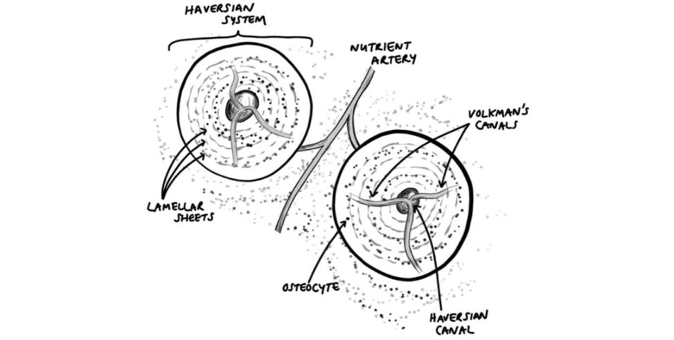
Candidates must know about the biological material we treat. It is simply inexcusable not to. One must know the functions, the structure, the.
With regards to cortical bone, one type of lamellar bone (with the other.
Figure 31.29 Bone (cortical).
It clearly shows the arrangement of haversian systems.
Within the haversian system there are central Haversian canals surrounded by concentric lamellar sheets, or rings, made of collagen. Within the lamellae sit osteocytes, which are connected by cannaliculi.
Overlying structure of nerve/muscle/tendon
All these structures can be drawn the same basically as they are all made up.
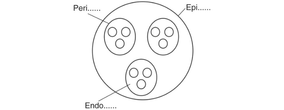
Figure 31.30 Overlying structure of nerve/muscle/tendon.
Nerve
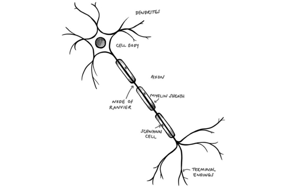
Figure 31.31 Nerve.
The structure of a standard axon connecting a cell body to terminal endings is a common starting point Figure 31.31).
Nerve cross-section (Figure 31.32)
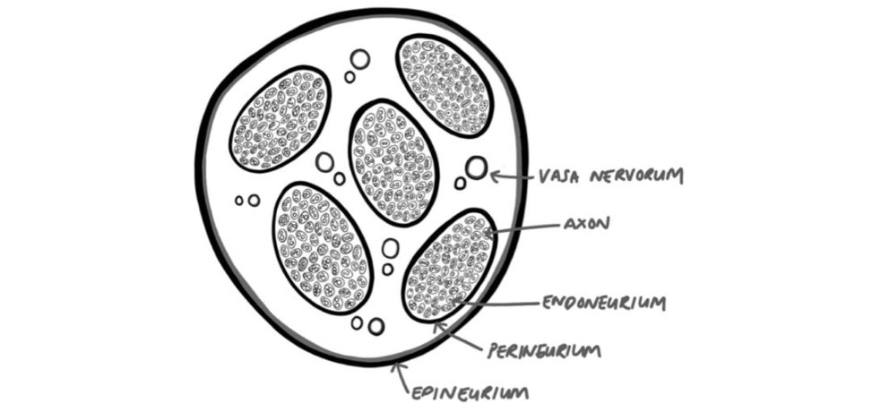
Figure 31.32 Nerve cross-section.
For a nerve this can be more specifically drawn as shown (Figure 31.32). The blood supply is both carried intrinsically within the endoneurium and extrinsically from the vasa nervorum.
Nerve action potential (Figure 31.33)
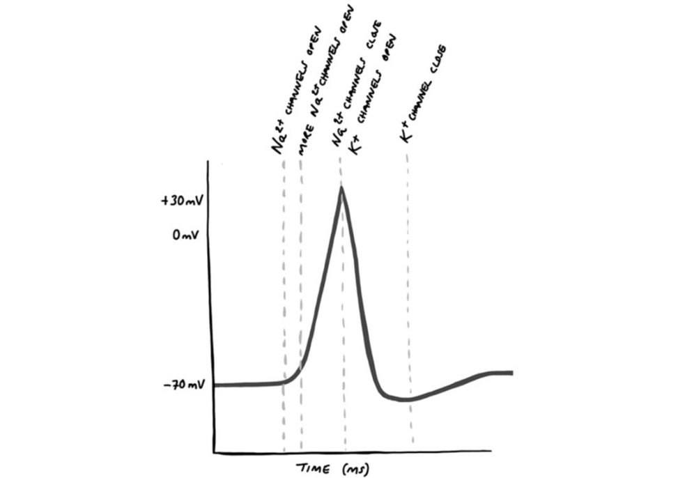
Figure 31.33 Nerve action potential.
It is a rapid depolarization across the membrane which then propagates along the neuron.
A threshold stimulus must first be reached (–55 mV) to trigger an action potential.
This propagates from cell to cell, causing more and
(a positively charged ion) into the cell causes the membrane potential to become positive. For cardiac muscle, the depolarization is halted temporarily by a plateau caused by calcium ions from the sarcoplasmic reticulum.
Nerve injuries (Figure 31.34)
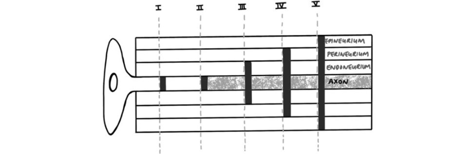
Figure 31.34 Nerve injuries.
Nerve injuries can be classified inaccordance with the mechanism of injury (crush,
Sunderland (1951).
The above illustration detailsS underland’s anatomical classification.
1: disruption to myelin/ischaemia to nerve – a.
2: axonal discontinuity – leads to Wallerian degeneration.
3: endoneurium damage (as well as axon) –.
4: perineurium damage (as well as endoneurium and.
5: total transection of nerve (epi-, peri-, endo- and.
NB: Wallerian degeneration – discovered by Waller in 1850, a.
Muscle (Figure 31.35)
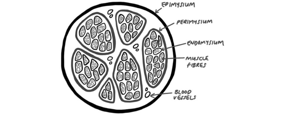
Figure 31.35 Muscle.
The external layer is the epimysium, the perimysium surrounds muscle fascicles (as it surrounds nerve fascicles) and the endomysium surrounds muscle fibres.
which themselves are made from sarcomeres containing actin and myosin.
Actin and myosin (Figure 31.36)
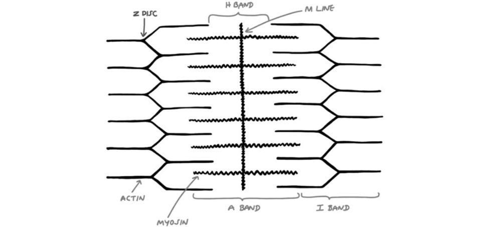
Figure 31.36 Actin and myosin .
Z disc – attachment between adjacent sarcomeres.
H band – only myosin filaments.
M line – connections between adjacent myosin filaments.
A band – both actin and myosin filaments.
I bands – only actin filaments (so-called as.
The sarcomere is the motor unit responsible for contraction in the muscle fibre.
Neuromuscular junction (Figure 31.37)
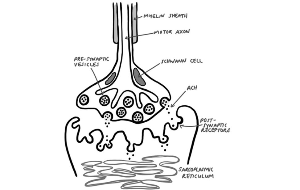
Figure 31.37 ACH, acetylcholine (neurotransmift er).
The candidate needs to.
When an action potential is delivered to a neuromuscular junction, acetylcholine is released from presynaptic vesicles across the synaptic cleft.
The myosin filaments then rotate to bindwith actin and ATPase activity permits conformational change in the actin which generates the sliding of the two filaments, causing muscle contraction.
Embryology – spinal development (Figure 31.38)
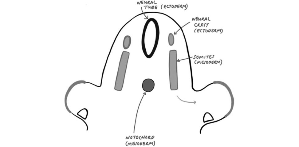
but FRCS candidates only need a basic understanding.
There are three germ layers
Ectoderm (‘outer’) – forms the skin and nerves.
Mesoderm (‘middle’) – forms muscles and cartilage.
Endoderm (‘inner’) – forms organs.
Figure 31.38 shows a cross-section of an embryo at around day 30. It shows.
Figure 31.38 Embryology – spinal development.
At day 25, a process called neurulation occurs where the notochord sends messengers to the neural tube (which starts on the outside of the embryo) to fold in on itself, forming a tube, in order to look like it does in Figure 31.38.
Following this, spinal development is controlled by the homeobox gene group. The notochord forms the anterior vertebral bodies and nucleus propulsus and the somites form the rest of the vertebral bodies and annulus fibrosis.
Embryology – limb bud development (Figure 31.39)
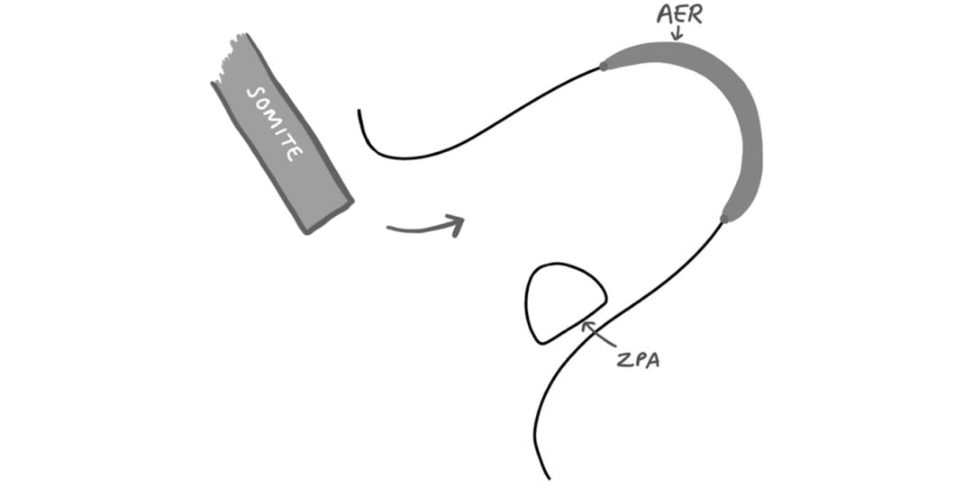
Figure 31.39 AER, apical ectodermal ridge; ZPA, zone of polarizing activity .
Limb development starts at around 4 weeks. At 8 weeks the limb buds rotate, which is.
There are three axes of growth:
1: Proximal to distal – controlled by the homeobox gene and performed by the AER.
2: Radial to ulna – controlled by the sonic hedgehog gene and performed by the ZPA.
3: Dorsal to ventral – controlled by the WnT gene and performed by the surface ectoderm.
The cartilage and muscle are derived from.
Implant science
Stress–strain curve (Figure 31.40)
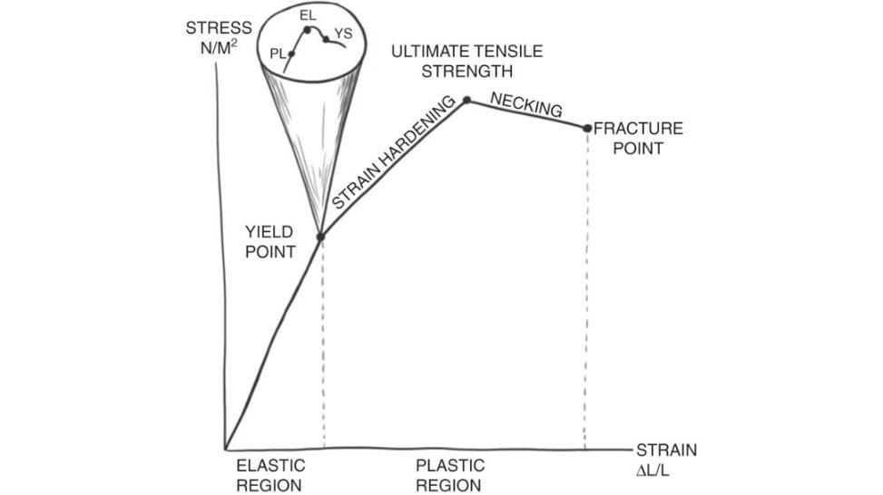
Figure 31.40 Stress–strain curve.
Stress–strain curves are commonplace in the FRCS. Start by drawing and labelling the two axes –stress (N/m2) and strain (change in length / original length).
In this region a specific stress–strain point reveals the material’s.
The yield point heralds the change from elastic to plastic region.
Proportionality limit – the highest point at which.
Elastic limit – the point at which the.
Yield stress – the amount of stress necessary.
This is where the grains in the metal dislocate and slip, paradoxically increasing the resistance to further strain.
Necking is where further strain occurs but in a relatively small area of the material, resulting in a decreasing surface area.
S-N curve (Figure 31.41)
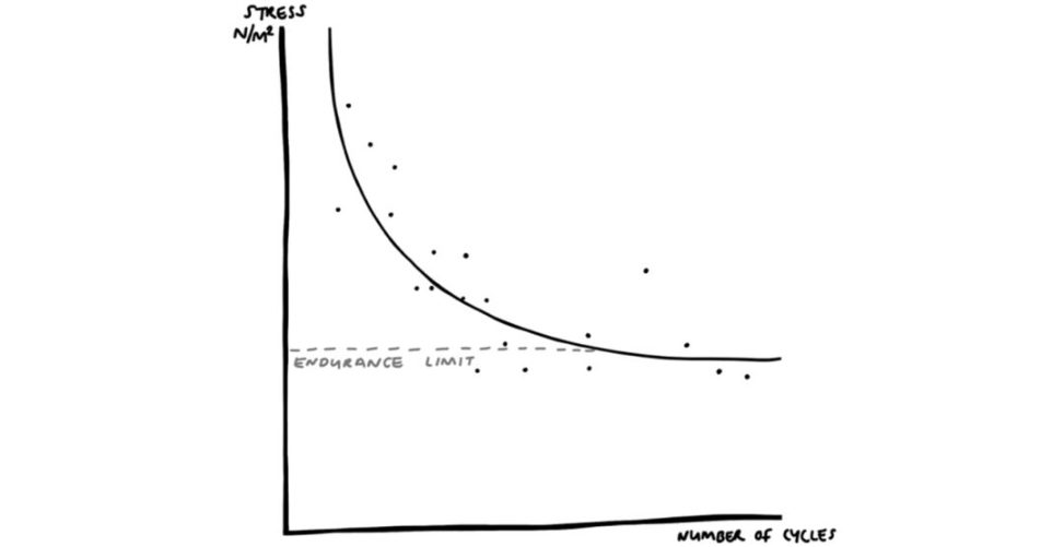
Figure 31.41 S-N curve.
The axes are first drawn with stress (N/m2) and number of cycles of stress application. The stress is always below that of the ultimate tensile stressor else the material would break on the first cycle of loading. A point is then made for each level of stress and the amount of times it can be applied before the material reaches fatigue failure and breaks.
Where the line plateaus out corresponds to 10 million cycles of loading for that level of stress without fracturing. This is called the endurance.
Viscoelastic behaviour (Figure 31.42)
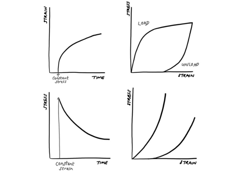
Figure 31.42 Viscoelastic behaviour.
Viscoelastic behaviour demonstrates time-dependen t deformation.
Creep – a change in strain over time, under a constant stress. This clinically translates.
Hysteresis – a different stress–strain relationship is seen between loading and unloading a material. This is often down to energy being lost (often.
Stress relaxation – a change in stress over time, under a constant strain. This can beseen when implanting uncemented femoral.
Time-dependent behaviour – this demonstrates a different stress–strain.
Time-dependent behaviour explains why low-energy injuries result.
Screws (Figure 31.43)
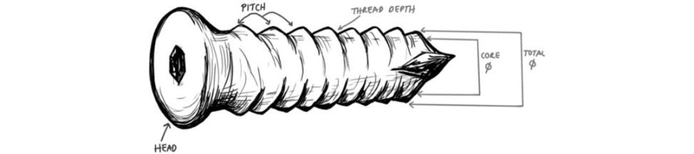
Figure 31.43 Screw.
Screws are a mechanical device used to convert torque (rotatory load) into linear motion. An FRCS candidate should be able to talk in depth about this common orthopaedic implant. The head can be of multiple shapes (hexagonal, star, cross-head, etc.) and on metalwork removal it is imperative to have the correct screwdriver. The pitchis the frequency of threads and defines the rate of linear motion per 360° turn. screw diameter, working length, locking screws, quality of bone) could all be discussed further in a viva scenario.
Tension band (Figure 31.44)
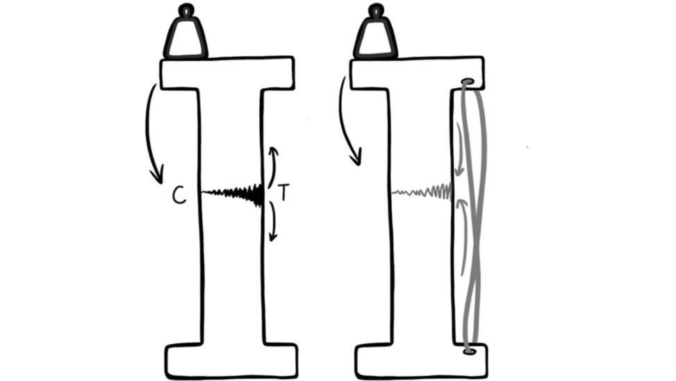
Candidates could be asked to explain the principle behind a tension band fixation (of an olecranon, for example). We found this easiest when utilizing an illustration such as the one in Figure 31.44.
side. Eccentric loading occurs incurved bones. By applying a tension band (in this case a figure-of-eight wire, similar to that seen in olecranon fracture fixation), the tensile forces from the eccentric load have now been converted to compressive forces. This can be done.
Figure 31.44 Tension band.
Cement zones (Figure 31.45)
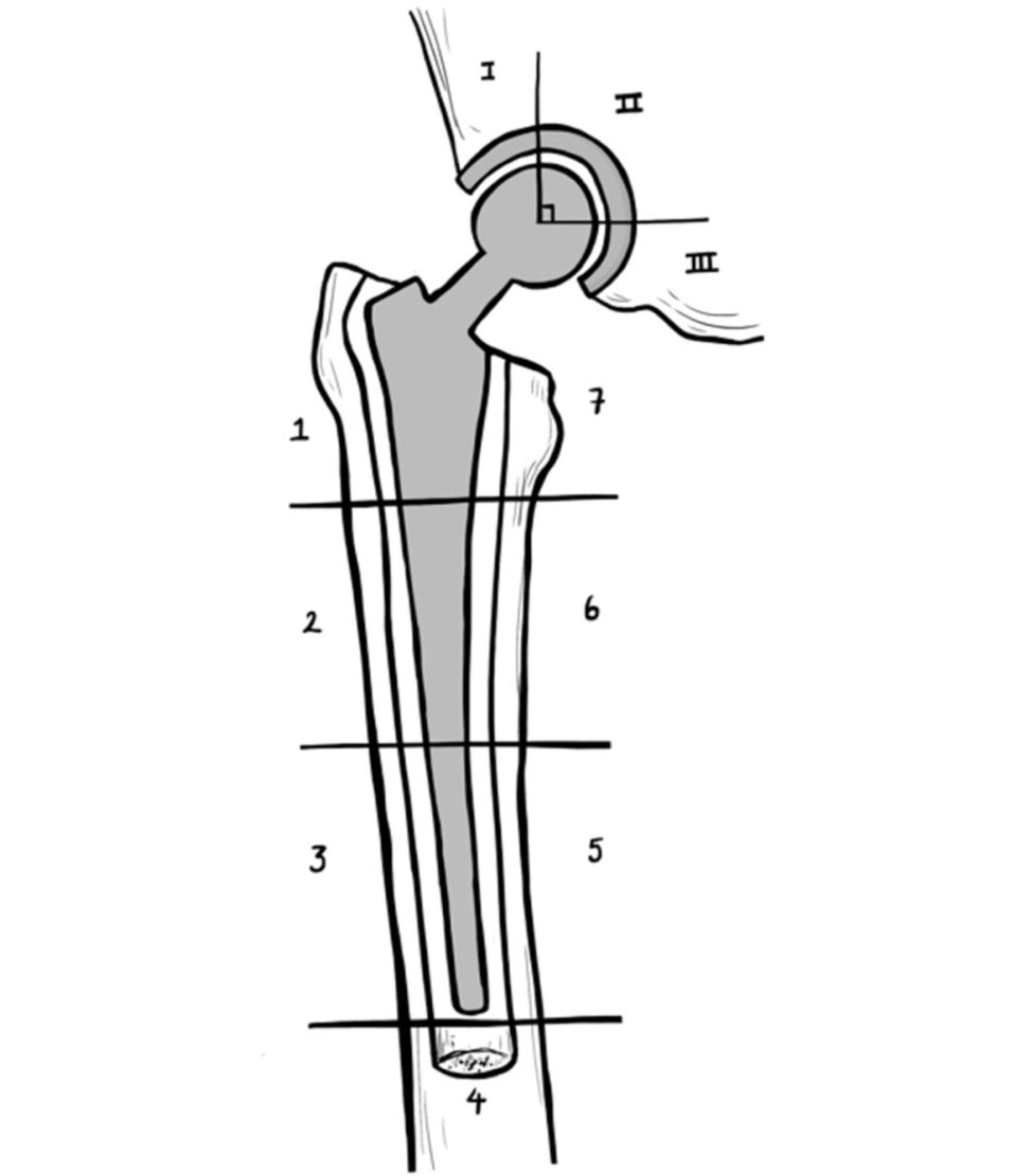
Figure 31.45 Cement zones.
Cemented total hips can be assessed
Charnley’s zones in the acetabulum.
Zones 1 and 7 are at the level of the greater trochanter and lesser trochanter, respectively Zone 4 is to the tip of the prosthesis but within the cement tail.
Zone 1 is the superior third, zone 2 is the middle third and zone 3 is the medial third.
Spinal pathology#
Spinal cord anatomy (Figure 31.46)
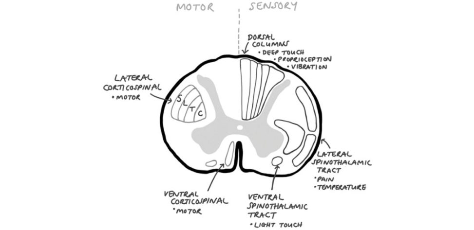
Figure 31.46 can be used not only to demonstrate both the anatomy of the spinal
Dorsal columns – deep touch, proprioception vibration.
These tracts cross at the spinal cord, explaining the clinical picture seen in
Brown–Sequard syndrome from a cord hemitransection where the ipsilateral side experiences loss of motor.
Ventral spinothalamic tracts – these carry light touch.
Figure 31.46 Spinal cord anatomy.
On the opposite side the motor tracts are shown:
Lateral corticospinal tracts – carry motor pathways distally. This explains why in central cord syndrome, the patient will retain more function in their lo wer limbs than their upper limbs.
Ventral corticospinal tracts – carry motor pathways distally.
Schematic (Figure 31.47)
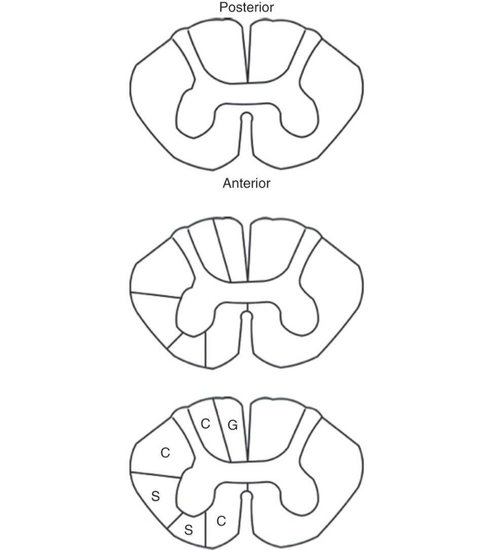
1. Draw a dumbell shape, and label anterior and posterior.
2. Draw a buft erfly within the dumbell shape and label it as ‘grey matter’ with anterior and posterior horn.
3. Divide the spinal cord into halves. On one half, split.
4. These are then labelled from posterior to anterior with ‘GCCSSC’. This is like the GCS ort heUK school exam GCSE.
Spinothalamic tract, anterior Spinothalamic and anterior Corticospinal tract.
Figure 31.47 Schematic drawing of spinal cord anatomy.
5. This is an important distinction to know as this determines the defects seen incertain spinal cord syndromes. looking from ground to cephalad) or with some imagination
ATLS (if on left side of c ord). This is important in the defects it causes in central.
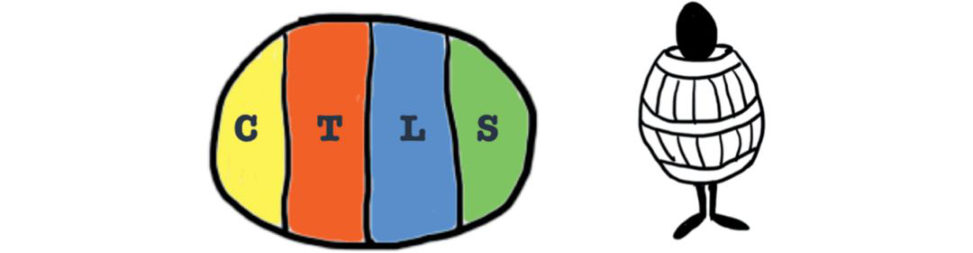
Figure 31.48 Topography of corticospinal tracts.
Vertebral disc (Figure 31.49)
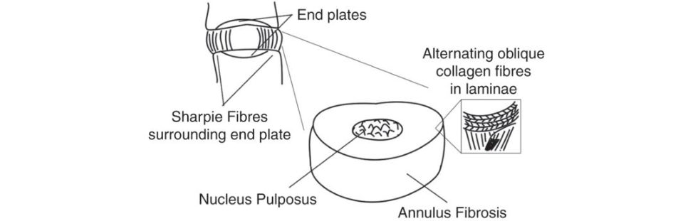
Intervertebral discs consist of an outer fibrous ring
(laminae) of fibrocartilage made up of type I collagen, which are obliquely orientated, alternating every layer.
The inner gel-like centre, the nucleus pulposus, is composed of type II collagen. This prevents the development of stress concentrations, which could cause damage to the underlying vertebrae or to their endplates.
Figure 31.49 Vertebral disc.
Prolapsed intervertebral discs (Figure 31.50)
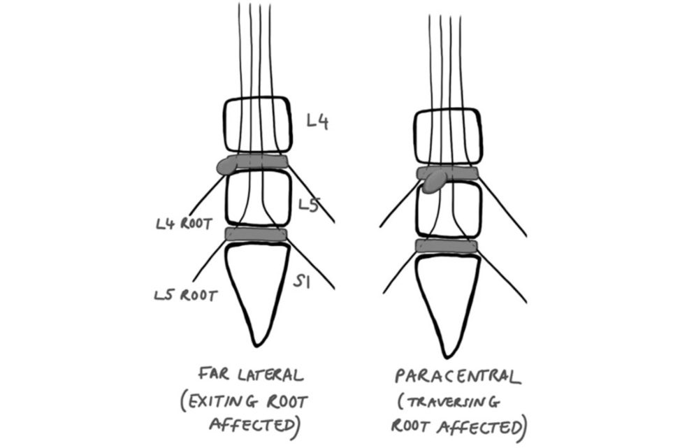
Figure 31.50 Prolapsed intervertebral discs.
Patients with nerveroot signs are often seen on the clinical day of the FRCS (Tr & Orth). It is important for the candidate to switily determine the.
At each lumbar level there is an exiting root and
L4 rootis exiting and the L5 rootis traversing.
A far lateral disc prolapse will affect the exiting root. Hence a far lateral L4/5 disc will cause L4 root signs. This.
L5) root, leaving the exiting L4 root free from compression.
Paediatrics#
Physis (Figure 31.51)
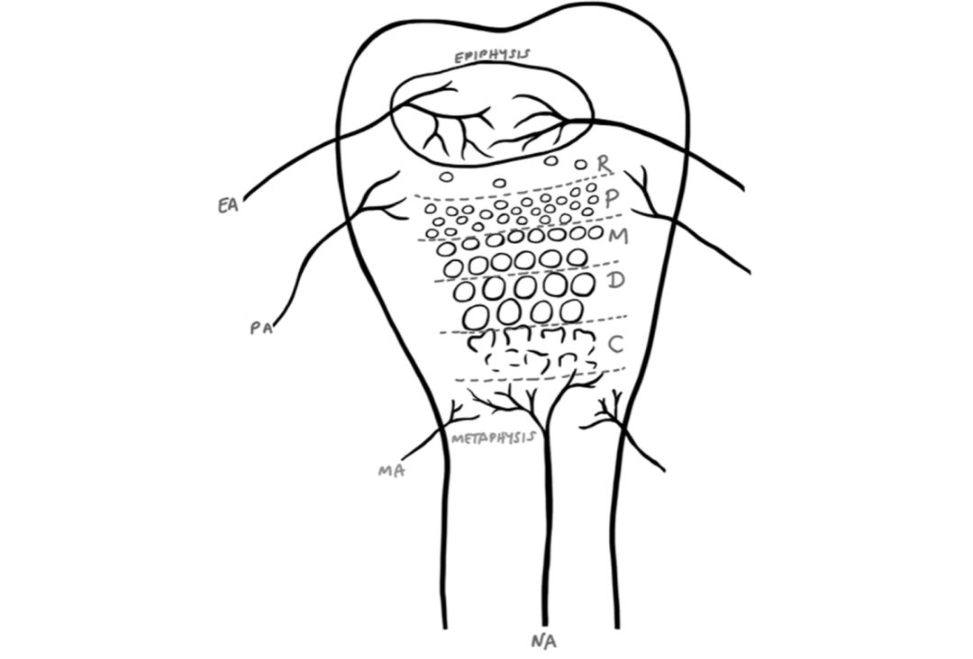
Figure 31.51 EA, epiphyseal artery; PA, perichondrial ring artery; MA, metaphyseal artery; NA, nutrient artery.
reserve zone, stores glycogen and lipids.
P, proliferative zone – proliferation of chondrocytes.
M, maturation z one – chondrocyte growth.
continued chondrocyte growth (×5 in size).
chondrocyte death permits calcification.
NB: hypertrophic zone includes maturation, degenerative and calcification.
One should be aware of the various diseases that can affect each zone of the physis, either from hypo- or hyperactivity , and the clinical manifestations.
Salenius and Vankka graph (Figure 31.52)
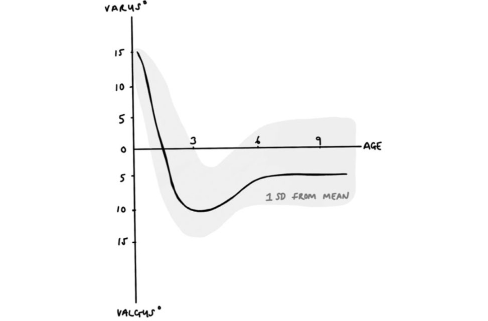
Figure 31.52 Salenius and Vankka graph.
Assessing limb deformities is commonplace in the paediatric clinical cases. Thus an understanding of normality.
reproduction of the above graph can be useful to further back your diagnosis of either physiological.
Essentially , newborns exhibit roughly 15° of varus which.
This progresses to 10° valgus at 3 years and resolves to adult values (5–7°) by 7 years of age.
Index
Page numbers followed by n refer to notes.
AAOS classification (acetabular defects) 30
127 accuracy 699 acetabular cup removal 33–34 acetabular lines 293 acetabulum bilateral fracture dislocation 303–304 classification of defects 30–31
DDH 51–55, 417 exposure in THA 432 fracture 299–301 fragment
395 achondroplasia 641–642 acidosis 308
ACL. See anterior cruciate ligament acromioclavicular joint (ACJ) dislocation 276–277
Adams’ forward bending test 150 adductor canal 722 blocks 568 adhesive wear 46–46
Blount’s disease 408, 411 scoliosis 149–151 adrenaline 444, 579, 581 advanced glycosylation end products 479–480 adverse reactions to metal debris (ARMD) 23–26, 44 age. See elderly patients air filters 613–614, 686 alendronate
WHO pain ladder 570–571 See also 581 713–723
Anderson and D’Alonzo classification 319–320, 654 trimalleolar 438–441 triplane 347–348 lateral ligament instability 96–98 surgical approaches anterolateral 242 posterolateral 263–264, 438–441 tarsal tunnel 448–449 ankle–brachial pressure index (ABPI) 222 ankylosing spondylitis 146–149 annulus fibrosus 490–491, 497 anterior cervical discectomy and fusion 313 anterior cord syndrome 316, 527 anterior cruciate ligament (ACL)
82–83 stress–strain curve 516 anterior drawer test 96 anterior talofibular ligament 96 antibiotics 612 arthroplasty prophylaxis 612 bites 288, 224 prosthetic joint infections 22–22 resistance 612
AO classification 324, 325–326
AORI classification 85 arch index 111
nails 183, 185, 186–187 plates 190–191
ARMD (adverse reactions to metal debris) 23–26, 44 arterial blood gases 256–257,
OA 175–179
RA 172–175, 178 foot 100–103 hand 363–364, 376–378
CMC thumb joint 364–366
MCP joints 291–292, 363, 364 hip
OA 34–38 septic arthritis 10–11
RA an dOA compared 174 wrist 362–363 See also osteoarthritis arthrodesis ankle 99
Lisfranc injuries 236
MCP joints 291–292 spine
C1/C2 320 cervical facet dislocation 313 lumbar interbody fusion 144, 656 arthroplasty ankle distraction 99 replacement 99–100 antibiotics 22–22
See total hip arthroplasty implant materials 554–555, 557, 605, 627–629 See also polyethylene knee total.
MCP joints 292, 364 postoperative analgesia 568–569 shoulder 160, 278 arthroscopy ankle 99 capsular release 163–164 elbow 175–177 knee 232, 724 chondrosarcoma 591–592 drawing 724 enchondroma 369–370, 648 structure/composition 466–467
AS IT GRIPS 3Cs 39 aseptic loosening 29,
ASIA scale 325, 330
Aspen collars 320 aspirin 378, 616 assessors (in exams) 4
ATLS protocols 294, 304–305 spinal trauma 311, 329
ATMIST 307
ATP (adenosine triphosphate) 501–502 atypical lipoma 596–597 autogratis 82
hip 38–45, 211, 648 in SUFE 382, 385 humeral head/neck 278
hangman’s fracture 321–322 odontoid peg fracture 321 axonotmesis 521–522 back braces 153–154 back pain disc disease 122
Baxter nerve 449 bell-shaped curves 689–690
nails 183, 185, 186–187 plates 190–191 benign tumours aneurysmal bone cyst
Berger flap technique 282 best-practice tariffs 213 bias
Bier’s blocks 573–575
Bigelow manoeuvre 214–215, 243 bioabsorbable materials 205 biofilms 260 biologic agents and surgery 102, 623 cartilage 477–479 femoral stems 546–547, 604–605 free-body diagrams 629–638 intervertebral discs 490
S-N curves 182, 608–609 tendon 516, 629 viscoelasticity 477
Young’s modulus 605, 619 See also stress and strain biopsy 130
CRPS 572 osteoporosis 611, 657–658
Paget’s disease 50–50 bites 287–288 290–292
Bleck classification 396 blood loss hypovolaemic shock 255–256, 720 humeral head 278 interrupted ankle fracture–dislocation 227–228 at fracture sites 259 knee dislocation 221–222 intervertebral discs 154–155
Blount’s disease 411
BOAST guidelines 222–223, 295 bone 452–469, 601–602 biomechanics 455, 461, 623 blood supply 456–457, 602 cell types 454–455, 462–464, 466, 601–602 collagen 457–458, 459–461, 642 densitometry 610, 611, 657–659 drawing 725 dysplasia 462 function 452 growth plates 467–469, 648, 734–735 healing 194–197, 259–260, 464–466, 609 in plate fixation 275 primary 196, 272, 464, 608 secondary 196–197, 464–465 stem cell therapy 648 See also non-union of fractures loss.
‘bone-healing organ’ 465–466
Boneloc cement 60, 551
Boston brace 153–154, 562 boundary lubrication 600
376 bowed legs (genu varum) 411, 693 boxer’s fracture 288–289 brachial artery 339, 714 brachioradialis 268 britile materials 622
Broström/modified Broström procedures 97
Brown–Sequard syndrome 316, 527
BSSH classification 372 bucket handle tears 72–73
Budapest criteria 572 bulbocavernosus reflex 318 bunions (hallux
Butler’s procedure 390 but iress plates 232
C-reactiv e protein (CRP) 399 calcaneal fracture 237–239 calcaneal osteotomy 106, 107 calcaneocavus foot 104 calcaneofibular ligament 96 calcaneovalgus foot, congenital 410–412 calcific tendonitis (shoulder) 164–165 calcium role in muscle contraction 500–501
Ewing’s sarcoma 595–596 osteosarcoma 593–594 spinal metastases 123, 167 discussing controversial issues 14–15, 465 tact 219 cannulated screws 200
Cannulok hip 57
Canterbury scale 675
scaphoid carpal tunnel anatomy 718 carpal tunnel syndrome 449–451, 674–676 carpometacarpal (CMC) joint (thumb) 364–366 cartilage See articular cartilage;
Caton–Deschamps method 87 caudae quina syndrome 138–142, 327–329
CAVE deformity (clubfoot) 391–395, 397 cavovarus foot 104 cavus foot 103–106 cefuroxime 612 cell savers 32 cement
709 central cord syndrome 314–316, 526–527 cephalic vein 444 cephalomedullary nails 245–247 ceramic implants
CoC arthroplasty 43, 44
CoP arthroplasty 43 liner removal 34
326–327 hangman’s fracture 321–322 odontoid peg fracture 321 prolapsed disc 154–157
RA 173, 378 spinal cord injuries 314–316, 526–527 spondylotic myelopathy 135–138 surgical approaches 316
Chance fracture 323–324
Charcot–Marie–Tooth (CMT) disease 104
Charnley THA implants 544, 546–547, 605
Charnley zones 732 chauffeur’s fracture 267–268 cheilectomy 118 chemical shift artefacts 657 chemotherapy 594, 724 chondrosarcoma 591–592 chronic (complex) regional pain syndrome (CRPS) 352
ACJ dislocation 276–277 fracture 275–276 clinical trials critical analysis 699–702 design 693–698 clopidogrel
CMAPs (compound muscle action potentials) 661–662
Cobb angle 150
Codman’s triangle 593
Coleman block test 104–105 collagen in bone 457–458, 459–460 osteogenesis imperfecta 460–461, 519 collagenase injections 373 common peroneal nerve 69, 65n compartment syndrome
723 complex (chronic) regional pain syndrome (CRPS) 352, 571–573 complex repetiv e discharges 670–671 compound muscle action potentials (CMAP s) 661–662
330 timing 294 trauma scans 256 wrist 285 computer navigation in TKA 542–543 conductance 525 conduction velocity 663–664 conduit gratis 530 confidence interval (CI) 699–701 conformity 542 congenital calcaneovalgus foot 410–412 congenital scoliosis 151–152 consent 252 contrast agents 655
175 coracoid osteotomy 444 corrosive wear 533 cortical bone 453–454, 609 corticospinal tracts 733 corticosteroids See steroids cotyloplasty 53 countersinking a screw 201–201, 205 cover-up test 406–407 crack propagation 182, 13–14 creep 477, 40 cross-bridge cycle 501
Crowe classification 52
CRP (C-reactiv e protein) 399 crystalloids 306
CT. See computed tomography cubital tunnel syndrome 676–677
DAIR procedure 20 damage control orthopaedics (DCO) 256–257,
DDH. See developmental dysplasia of the hip
De Quervain’s tenosynovitis 449 debridement arthritis 99, 269 pseudotumours 26 degenerative spondylolisthesis 143
Denis Browne boots 395
Denis classification 303 denosumab 595
in adults 51–55 in children 417–426
DEXA (dual-energy X-ray absorptiome try) scans 610, 674–679 statistics 698–699 dial test 71 diathermy 683 diffuse idiopathic skeletal hyperostosis (DISH) 147
See intervertebral discs disease modifying anft-rheuma toid drugs (DMARDs) 102, 364, 378 dislocation
ACJ 276–277 cervical facet 311–314 hip 214–216
PE spacer 79–80 radius
Galeazzi fracture 272–273
Monteggia fracture 270–271, 335 shoulder 265–266 wrist 281–282,
378 distal metatarsal articular angle 113–115 distal radioulnar joint dislocation (Galeazzi fracture) 272–273 distraction arthroplasty 99 divot sign 23
DNA, structure 639
Dorr grade of femur 35 dorsal root 714 651–652
Doyle’s classification 287
Drehmann sign 379
Duchenne muscular dystrophy 645–646 ductile materials 622, 625
685 dynamic condylar screw (DCS) implants 206–207 dynamic hip screw (DHS) implants 206
‘early appropriate care’ 308
Eaton and Litiler classification 365 effective joint space
Eikenella corrodens 291 elastic limit 619 elastic materials 627 elastic nails 333–335, 342–343 elastin 510 elastohydrodynamic
385–390 free-body diagram 630–632 osteoarthritis 175–179 osteochondritis dissec ans 170–172 paediatrics 333–339, 180 posterolateral rotatory instability 179–180 rheumatoid arthritis 172–175, 178 tennis elbow 168–170 ulnar nerve compression 676–677 elderly patients acetabular fracture 300–301 cartilage 37, 609–612 electromyography (EMG) 662
Elson’s test 376 embolization pelvic injuries 306 spinal metastases 131 embryology 728 emergency surgery ankle reduction 415 caudae quina syndrome 141, 668 enhanced recovery after surgery (ERAS) 568–569
Enneking staging system 587 entrainment 686
Erb’s point 715 erosive wear 533 ethics of surgeons carryingS.
Ewing’s sarcoma 595–596 examiners 3–4
604–605 explant acetabular cup removal system 34 explosion injuries 255–257 extended trochanteric osteotomy (ETO) 432 extensor carpi radialis brevis (ERCB) 169 extensor digitorum communis (EDC) 169 extensor pollicis longus (EPL) tendon 354–356
bone 459 cartilage 471, 472–475 meniscus 72, 487
F waves 666–667 falls caudae quina syndrome 138 in the elderly 135, 212, 300–301 from a height 237, 301–302 fasciculations
S-N curves 182, 624–625, 729
FATSAT images 656 femoral locking plates 219, 432 block 568 femoral stems
Cannulok 57
Charnley 544, 546–547, 605
Exeter 17, 35–36, 604–605 failure 45, 547, 606 femoral triangle 720–721 femoral varus derotation
DD Hin adults 51–55 in children 417–426, 698–699 dislocation 214–216, 243–245
DCS/DHS implants 206–207, 211 inter-trochanteric 245–247 near physis 343–345 neck, in the elderly 212–214, 233–234 neck, plus shaft 217–218 neck,
Legg Calvé Perthes disease 399–404 osteonecrosis 38–45, 211,
Paget’s disease 49 shape (Dorr grade) 35
SUFE 384–385 and TK Ain valgus knee 68–69 fever 127
Ficat classification 40 fight
Boutonnière deformity 363, 376 enchondroma 369–370
366–368 inability to extend MCP joints 174 mallet finger 286–287 swanneck deformity 363, 562 flexor digitorum longus (FDL) tendon transfer 111–112 flexor digitorum profundus (FDP) tendon 283–284, 368 flexor digitorum superficialis (FDS) 283 flexor digitorum superficialis (FDS) tendon 368, 305–306 fluid-film lubrication 600 foot arthritis 100–103 calcaneal fracture 237–239 calcaneovalgus foot 410–412 cavus foot 103–106 clubfoot 391–395
Lisfranc tarsometatarsal fracture dislocation 234–236 metatarsus adductus 395–397 navicular fracture 250–252 overlapping fitih toe 390 postoperative foot drop 58 prosthetics
Galeazzi 272–273
Monteggia 270–271, 333–335 surgical approaches 352–353, 433–436, 717 forest plots 702–704 fracture gaps 194–195, 464 fracture point of a material 618, 622 fractures bone
FRAX score 658 free body diagrams (FBDs) 629–638
551 freting w ear 46 frozen shoulder 162–164 functional bracing 562 funnel plots 705 gabapentin 570
Galeazzi fracture 272–273 gallium imaging 660 195
Garden Alignment Index 210
Gartland classification 387 gastrocnemius lengthening 106 gate theory of 575 581–584 639–640
X-linked dominant 643–645
X-linked recessive 645–646 genitourinary system caudae quina syndrome 327–328 trauma
Bier’s blocks in A&E 574–575 breakdown in infection
GRAFO orthosis 561–562 gratis bone.
Gram stain 684 grind test 366
Gross and associates classification 30–30 ground substance 510 growth factors 468–469
Gruen zones 45, 535–536, 732
Gustilo ’s classification 19
Guyon’s (ulnar) canal 718
112–116 hallux valgus interphalangeus angle 114 hallux varus 115–116 halo jackets/vests 320, 312 halogenated anaesthetics 583 hammer finger (mallet finger) 286–287 hamstring gratis 82, 376–378 thumb CMC joint 364–366 boxer’s fracture 288–289 carpal tunnel syndrome 449–451
Dupuytren’s disease 163, 371–373 enchondroma 369–370
EPL tendon rupture 354–356, 449
366–368 flexor tendon injuries 368–369 inability to extend MCP joints 174 mallet finger 286–287 and RA elbow 173
UCL injury (thumb) 375–376 307
Hardinge approach 36, 430–431, 432 hardness 618–619
Harris and Barrack scale 28
Hartofilakidis classification 52
Haruguchi classification 263
Hatirup and Johnson grading system 117
Haversian system 454, 462–464
Hawkins’ sign 242–243 health 651–652
111 hemiarthroplasty 212–213 hemivertebrae 151
717 hereditary sensorimotor neuropathy (HSMN) 104
Herring classification 400–401 heterogeneity 703
Hibb’s angle 105–106 high tibial osteotomy (HTO) 76–77, 111 hip anatomy 720–721 ankylosing spondylitis 148 arthroplasty hemiarthroplasty 212–213
MoM resurfacing 23–27, 43 total. See total hip
DD Hin adults 51–55 in children 417–426, 698–699
DDH 52, 417–426 fracture dislocation 214–216 fractures
DHS 206, 211 in the elderly 212–214, 233–234, 245–247 inter-trochanteric 245–247 periprosthetic
Legg Calvé Perthes disease 399–404 osteoarthritis 34–38 osteonecrosis
Paget’s disease 48 pain 25 29
Paget’s disease 50
PJI 18 periprosthetic infection 17–23 septic arthritis 10–11,
SUFE 384–385 surgical approaches 11, 245 for THA 36, 429–432 zone of safety 419
Hoffmann’s sign (H reflex) 667–668
Hooke’s law 618 hoop stresses
IM nails 188, 193 intervertebral discs 495–496 meniscus
Hounsfield units 654
Hueter–Volkman law 455 human bites 290–292 humeral spaces 715 humerus anatomy 715–716 distal fractures comminuted 268–270 lateral condylar 335–337 supracondylar 337–339
Hunter’s (adductor) canal 722 blocks 568
HXLPE (highly cross-linked PE) 26, 556 hyaluronic acid 480 hydrodynamic lubrication 600 hydroxyapatit e-coated implants 628 hypophosphataemic rickets 644 hypotension after pelvic injury 297, 331 hysteresis 477
IL-1 (interleukin-1) 38
IL-6 (interleukin-6) 18, 51 iliac oblique view (Judet)
IM (intramedullary) nails. See nails implants failure. See wear materials 554–555, 557, 605 See also specific types induction agents 582 infection control 22, 612, 684–685 sterilization 681 in theatre 22, 614, 685–687 infections bites 287–288, 290–292 discitis 122–127, 154–155, 498 flexor sheath 287–288,
X-linked dominant 643–645
X-linked recessive 645–646 insertional activity on EMG 668 intention-t o-treat (ITT) 698 interleukin-1 (IL-1) 38 interleukin-6 (IL-6) 18, 51 interlocking screws 188–189, 199 intermetatarsal angle 113–115 interphalangeal joint (finger). See distal interphalangeal joint; proximal interphalangeal joint interphalangeal joint (toe) 117 intervertebral discs 489 age-related changes 154, 492, 494–495 blood supply 154–155, 492 discitis 122–127, 154–155, 498 function 490, 491–492, 494, 495–497
Johnson and Strom classification 109 joint aspiration 18–19
Judet and Letournel classification 300
Judet view 298
K-wires ankle injuries 417 upper limb injuries 333,
Kanavel’s signs 287
Kaplan–Meier survival curve 709
Kaplan’s line 451
Keller’s arthroplasty 118
Kerboull necrotic angle 41
Kessler technique 369 ketamine 582 kidney disease 378, 655
Kienbock’s disease 373–375
See total knee arthroplasty arthroplasty, unicondylar 77–78 dislocated spacer 79–80 below-knee prostheses 562–565 dislocation 86–88, 220–222, 436–438 high tibial osteotomy 76–77, 78–79, 92–93 ligaments 70
ACL 71, 82–83, 485, 489, 516 in dislocation
MPFL 87, 88
PCL 71, 80–82 meniscus. See meniscus paediatric injuries 83, 345–347
PLC injury 70–71, 608–609 tibial tubercle fracture 345–347 knot of Henry 111–112
Kocher–Langenbeck approach 215, 245 posterior 295–296
Kocher’s criteria 10–11, 433 kyphoplasty 131 kyphosis 154 lactate 256–257, 685–686 laminectomy 137 laminoforaminotomy 157
Langenskiold classification 410 large fragment screws 201
Larmor equation 656
LARS reconstruction 277 lateral collateral ligament (LCL) 179 lateral condylar fracture 335–337 lateral epicondylitis tennis elbow) 168–170 lateral meniscus 72, 482–483,
Leadbetter technique 210
Leddy and Packer classification 283 leg length discrepancy
Legg Calvé Perthes disease (LCPD) 399–404 levels of
Levine and Edwards classification 322 levo-bupivacaine 580
Lewis and Rorabeck classification 219
Lichtman classification 374 lidocaine 580 ligaments 509–521 ankle 96–98 attachment to bone
LARS reconstruction 277 ligamentum flavum 516 stem cell therapy 648 stress–strain curves 515–516, 520, 623, 629 structure and function 509–510, 512, 514,
Lisfranc tarsometatarsal fracture dislocation 234–236 local anaesthesia 579–581 locking screws
Loder classification 382 lower limb anatomy 70, 735 compartment syndrome 224, 556 nerve conduction studies 677–679 prostheses 562–565 trauma 209
VTE prophylaxis 615–616 See also ankle; femur; foot; hip; knee; tibia lubrication in synovial joints 599–601 lumbago
L3–L5 burst fractures 318
L5 transverse process fracture 297
L5/S1 radiculopathy 677–679 metastases 128, 129
MRI 655
Paget’s disease 49 spondylolisthesis 142–146 See also thoracolumbar
Kienbock’s disease 373–375 transscaphoid perilunate fracture–dislocation 356–358 lung
Mafucci’s syndrome 370, 589 magic angle artefact 657
BMES 62 caudae quina syndrome 140–141
MARS 44
Kienbock’s disease 374 osteosarcoma 593 principles 654–657 spine 655 aneurysmal bone cyst 133 cervical disc prolapse 156 cervical facet dislocation 312–314 cervical spondylotic myelopathy 137 discitis 123, 125 metastases 129, 131 scoliosis
Masquelet technique 260 matrix glycoproteins 473–474 matrix metalloproteinases
Maudley’s test 170
Mayfield classification 356
McKellop’s classification 538
McPherson staging 74
MCPJ. See metacarpophalangeal joint
296 medial collateral ligament (MCL) 179 medial meniscus 72, 489 bucket handle tear 72–73 meniscectomy 483–484 medial patellofemoral ligament (MPFL) 87, 88 median nerve 253–254, 527–528 carpal tunnel syndrome 449–450, 514 meta-analysis 702–706 metabolic acidosis 308 metacarpal fracture (boxer’s fracture) 288–289 metacarpophalangeal joint (MCPJ)
inability to extend 174 infections 288, 290–291
OA 291–292
RA 363, 375 metal artifact reduction sequence (MAR S) 44 metal AVN rods 42
THA 44–44 metals 554–555 and MRI 655–656, 657 stress–strain curves 617, 621, 629 See also stainless steel titanium
arthritis 100–103 hallux rigidus 117–119 hallux valgus 100
Meyerding’s grading system 142
MHRA (Medicines and Healthcare Products Regulatory Agency) 24
Michon classification 368 mini fragment screws 201
Mitchell osteotomy 115
Modified New York Criteria 147
MoM (metal on metal) hips resurfacing 23–27, 43
THA 44–44 moment of inertias tiffness/rigidity)
nails 183–184, 185, 186–188 plates 190–191
Monteggia fracture dislocation 270–271, 333–335 motor endplates 525–526,
MRI. See magnetic resonance imaging
MRSA (methicillin-resistant S. aureus) 612, 684–685
MSIS definition of P JI 20
MTPJ. See metatarsophalangeal joint multiple injuries in the elderly 248 explosions 255–257 falls from height 237, 301–302 osteogenesis imperfecta 340 pelvic 306, 307 muramyl tripeptide 594 muscle 498 contraction 499–503, 505–509, 526 drawing 727–728 electromyography 662, 668–674 structure 498–499, 503–505 types 502–503, 504 muscle relaxants 582–583 muscular dystrophy 645–646 musculocutaneous nerve 444 myasthenia gravis 526 myeloma
‘Napoleon Hat’ sign 142 navicular fracture 250–252 neck. See cervical spine necking 621 needle biopsy 586–587 negative predictive
L5/S1 radiculopathy 677–679 lower limb 679 lower trunk brachial plexopathy 677 and type of nerve injury 521, 726–727 local anaesthetic mode of action 579–580 pain pathways 575
Wallerian degeneration 521, 526, 528 neurofibromatosis 152–154 neurogenic shock 317, 330–331 neuroma 653 neuromuscular blocking agents
‘NHS working environment’ 64n
575–579 See also pain non-normal distribution 692–693 non-ossifying fibroma 590–591 non-union of fractures clavicle 276 hip 246 humerus 275 odontoid peg 320 scaphoid 285–286
NSAIDs 378, 570 nuclear medicine imaging 63n, 659–661 bone marrow oedema 62–62 nucleus
OA. See osteoarthritis obesity 67, 77, 455 obturator oblique view (Judet) 298 occupational safety 651–652
Ollier’s disease 370, 589 oncology aneurysmal bone cyst 132–135, 594–595
127–132 non-ossifying fibroma 590–591 osteochondroma 587–588 osteosarcoma 593–594 staging and grading 130, 587 open book pelvis 304–306 open fractures at site of a bone tumour 591–592 femur 217 humerus 268 tibia 222–224, 685 infection control 22, definition 452 orthodromic action potential 664–665 orthotics 559–562 for bowed legs 409 for flafooot 111
OSCAR system 33 osteoarthritis (O A)
ankle 98–100 compared with RA 174 elbow 175–179
MCP joints 291–292 non-surgical treatments 67, 724 stem cell therapy 648 thumb CMC joint 364–366 osteoblastic jumping distance 260 osteoblastic regulators 47, 534 See also RANKL osteoblasts 455, 466 osteochondral autograft transfer system 477 osteochondral gratis 100 osteochondritis dissec ans 170–172 osteochondroma 587–588 osteoclastic cuting c ones 196, 642 osteolysis 47–48, 599 classification 29–31
Paget’s disease 51 revision knee replacement 83–86 osteon
hip 38–45, 211, 648 in SUFE 382, 385
DEX Ascans 610, 611, 657–659
FRAX score 658
ITOH 61–63
466 osteoprotegerin (OPG) 48 osteosarcoma 593–594 osteotomy ankylosing spondylitis 148 calcaneal 106, 111 coracoid 444 extended trochanteric 432 hallux valgus 115–116 high tibial 76–77
Paget’s disease 50 shortening of the femur 54 osteotropic factors 459 outcome measures 697
P values 696, 700–701
Padua grade (ulnar nerve compression) 677
Padua scale (carpal tunnel syndrome) 675 paediatrics 379
Blount’s disease 411 bowed legs 411, 735 discitis
397 congenital calcaneovalgus foot 410–412 congenital vertical talus 410 metatarsus adductus 395–397 overlapping fitih toe 390 forearm 333–335
DDH 417–426, 698–699
LCPD 399–404 septic arthritis 10–11, 399, 432–433
SUFE 384–385 knee
ACL injury 83 tibial tubercle fracture 345–347 limps 398–404 neurofibromatosis 152–154 osteogenesis imperfecta 340 rheumatoid arthritis 173 rickets 644 scoliosis 149–151
Paget’s disease 48–51 pain acute vs. chronic 568 analgesia
WHO pain ladder 570–571 See also anaesthesia assessment
CRPS 352, 571–573 definition 567 exam questions 567 knee 88–91 mechanisms 572
Paprosky classification (acetabular defects) 30
Paprosky classification femoral defects) 29, 30 paracetamol 570
Park Harris growth arrest lines 340
Pasteurella multocida 288 patellar instability 86–88 patellofemoral maltracking
PCL (posterior cruciate ligament) 71, 80–82
PD weighted images 656
PE. See polyethylene pedigree charts 640, 641, 643, 644, 645, 646, 647 pelvic anatomy 293, 297–298 pelvic binders 305, 308–309 pelvic fractures 293 acetabulum 299–301, 303–304 and bilateral dislocation 303–304 classification 297 lateral compression 307–310 open book 304–306 posterior wall 293–297 unstable 301–303 vertical shear 297–299 pelvic incidence 144–145
Perren’s strain theory 194, 465, 609 pes cavus
See growth plates pigeon toe (metatarsus adductus) 395–397 pilon fracture 227–229, 239–243 pinning in situ (PIS) 382–383, 384–385
PIPJ. See proximal interphalangeal joint (PIPJ)
Pipkin classification 215
Pirani score 391–394 piriformis 430 piriformis entry point 217 pivot-shift t est 180 plantar fascia release 106 plastic deformation 618, 620–621 plastic orthotics 560–561 platelet-rich plasma (PRP) injections 99, 275 clavicle 276 design 207–208 elbow 269–270 femoral locking plates 219
PLC (posterolateral corner) 70–71, 222
PMMA (polymethylmethacrylate) 605, 187 polyethylene (PE) 544
CoP arthroplasty 43
HXLPE 26, 556 liner removal 34 manufacture 48–48, 556 sterilization 539, 556 wear/failure 47, 555 polymethylmethacrylate (PMMA) 605
438 popliteal fossa 437–438 popliteal vein 438 positive predictive value (PPV) 699 positive sharp waves 668–670 positron emission tomography (PET) 64n, 659 post-tourniquet syndrome 682 post-traumatic arthritis 98, 80–82 posterior distal tibiofibular ligament 96 posterior interosseous nerve (PIN) 173, 222 postsynaptic membrane 526 power (statistics) 696–697 prilocaine 573–574 procalcitonin (PCT) 399
PROMs (patien t-reported outcome measures) 710
Propionibacterium acnes 19 propofol 582 proportionality limit 619 proprioception 135, 50 proximal femoral replacement 58 proximal interphalangeal joint (PIPJ)
Boutonnière deformity 363, 376 swanneck deformity 363
Punneft squares 640, 642, 643, 645 pyrexia 127 quadrangular space
RA. See rheumatoid arthritis radial artery 435 radial nerve 435 radiofrequency ablation 683 radioimaging 659–661 radiology 650–661
Galeazzi fracture 272–273
Monteggia fracture dislocation 270–271, 335 styloid fracture 267–268 surgical
RANKL (receptor activator of nuclear factor-κβ ligand) 47, 141–142 refractory period 525 regional anaesthesia 573–575, 307–308 reverse pivot shift t est 71 reverse self-cuting screws 205 reverse shoulder arthroplasty 167, 9–14 rheumatoid arthritis (RA)
compared with OA 174 elbow 172–175, 178 foot 100–103 hand 363–364, 376–378 medications 102, 364, 378
Risser grading 149–150
RNA, structure 639
Robert’s view of the thumb 364
ROC (receiver operating characteristic) 699
Romberg’s test 135 ropivacaine 580 rotator cuff tears
Ruedi–Allgower classification 240–241
S-N curves (stress cycle curves) 182, 624–625, 729
SACH (solid ankle cushioned heel) prosthesis 564 sacroiliac
Salenius and Vankka graph 405–406, 735
Salter Harris classification 467 sample size 696–697
Sanders classification 237–238
Sangeorzan classification 250–251 sarcoma chondrosarcoma 591–592
Ewing’s 595–596 osteosarcoma 593–594 sarcomere 498 scaphoid non-union of fractures 285–286, 360–361, 362 proximal pole fracture 284–286,
Schatzker classification 224
Schenk classification 221
Scho tiles point 88 sciatic nerve
DDH 53, 54 foot drop 58 hip dislocation 215–216, 243
THA 36, 432 scintigraphy technetium bone sc ans)
Boston brace 562 idiopathic 149–151 non-idiopathic 150, 151–154
DCS/DHS implants 206–207, 203 pull-out 195, 205 scrubbing up 687 seatbelt injuries 323–324
Seddon classification 529–530 self-tapping screws 205 sensitivity 698, 699 sensory nerve action potentials (SNAP s) 661, 665 sepsis 123 septic arthritis 10–11
ACJ dislocation 276–277 anatomy 444–445 arthroplasty 160, 656 subacromial impingement 164–167 surgical approaches 443–445 tuberculosis 158–160 sickle cell disease 643
Singh and Maini index 659 659
SIRS (systemic inflammatory response syndrome) 123, Paget’s disease 49 slipped upper femoral epiphysis (SUFE) 384–385 sloft ed nails 188 small fragment screws 201
Smith Peterson approach 211, 433, 721
SNAC wrist 362–363
SNAPs (sensory nerve action potentials) 661, 662, 665
SOCRATES 567 soleus bridge 447
Southwick angle 381 specificity 698, 699
SPECT (single photon emission tomography) 659 spilled teacup sign 358 spinal anaesthesia 585 spinal arteries 154 spinal shock 312, 317–318, 331 spine 121–122 anatomy 154, 489–490, 732–733 aneurysmal bone cyst 132–135
Chance 323–324 hangman’s 321–322
L5 transverse process 297 odontoid peg 321 thoracolumbar burst 316–319 free-body diagram 636–638 function 490, 497 fusion.
L5/S1 radiculopathy 677–679 ligamentum flavum 516 metastatic disease 123
331 stem cell therapy 648 stenosis 49, 328 surgical approaches (cervical) 316, 441–443 trauma assessment 329–331 spiral cords 372 splintage 184 spondylolisthesis 142–146
Spurling’s sign 155 staging of tumours 130, 587 stainless steel 343, 546,
Stanmore THA implants 27–28
Staphylococcus aureus
699–702 diagnostic tests 698–699 distribution 689–693 meta-analysis 702–706 outcome measures 697, 710–711 survival analysis 706–710 types of data/tests 690–691
Steel’s blanch sign 380
Steinberg classification 40 stem cells 42, 646–649
Stener lesion 375 sterilization 681 of PE 539, 546, 556 steroids elbow injections 170 joint infections 159–160 osteonecrosis 39 shoulder injections 159–160, 162,
STIR images 656 strain 617 in fracture healing 194, 465, 609 See also stress and strain strain hardening 621–622 strength 618, 626 stress corrosion 195 stress relaxation 477, 478, 626, 627 stress risers 182, 195 stress and strain 617–629, 729–731
Stulberg classification (modified) 401, 404 subacromial impingement 164–167 subtalar joint 109
Sunderland classification 529–530, 726–727 superficial peroneal nerve 242 superficial radial nerve 435 superior gluteal nerve 432 supervisors 13 sural nerve 440 surgical approaches 429 ankle anterolateral 242 posterolateral 263–264, 717 hand/fingers 355, 245 for THA 36, 681 surgical timeouts 435 survival analysis 706–710 sutures 682–683 suxamethonium 582–583 swanneck deformity 363, 376–378 synovial cyst (ganglion) 370–371 synovial fluid 599 aspiration in diagnosis of P JI 18–19 systemic inflammatory response syndrome (SIRS) 123
T score 610, 657
T1/T2-weighted images 654–655 talar fracture 241, 242–243, 654 talar tilt t
AVN rods 42, 43 tapping 195, 201 self-tapping
TE (timet o echo) 656 technetium bone sc ans 62–62, 659 telescopic rods 340 tendoachillis tenotomy 395 tendons attachment to bone 518 blood supply 517 healing 513–514 magic angle artefact 657 stem cell therapy 648 stress–strain curves 516
366–368 tensegrity architecture 474–475 tension banding 731
THA. See total hip arthroplasty thigh, anatomy 722 thiopentone 582 thoracic
Chance fracture 323–324 incomplete cord injuries 325–326 three-point pressure principle 561
CMC joint arthritis 364–366
EPL tendon rupture 354–356, 449 trigger thumb 397–398
UCL injury 375–376 tibia
347–348 non-union 257–260 open fractures 222–224, 256–257 periprosthetic (ankle) fracture 248–249 posterior malleolus fracture 263–264, 608–609 spiral fractures 183 tibial nails 193 tibial nerve 438, 449 tibial osteotomy (HTO) 76–77, 110 tibiofemoral angle (TFA) 405–406, 735 timing of C T scans 294 timing of surgery acute spinal cord injuries 316 ankle fractures 261–262, 415 caudae quina syndrome 141
TKA. See total knee arthroplasty
TLICS scale 325 toes complications of VF Gs 41 after FDL transfer 111–112
187 total ankle replacement 99–100 total elbow replacement (TER) 173, 177–178 total hip arthroplasty (THA)
for acetabular fracture 301 bone scans 660–661 cement mantle failure 58–61, 548–551 cemented
605 compared with hip resurfacing 27
DDH 53–54
625 femoral head size 556–557, 599 femoral neck fracture 213
604–605 lubrication 600 metal on metal 44–44 osteoarthritis 35–37 osteonecrosis 43–45
Paget’s disease 50–51 periprosthetic 55–58 17–23
DDH 54 periprosthetic fracture 56–58
PJI 21–22, 26–26
31–34 squeaking 43 stem biomechanics 546–547, 604–605 surgical approaches 36
598–599 total intravenous anaesthesia (TIVA) 583–584 total knee arthroplasty (TKA)
computer navigation 542–543 after HTO 78–79 implant design 85–86, 541–542, 543 implant materials 554–555 lubrication 600 pain following 88–91 periprosthetic infection
VTE prophylaxis 615–616 wear/failure 534, 539–544, 552, 625 toughness 618, 619, 627 tourniquet paralysis syndrome 682 tourniquets 573–574, 682 traction cervical 157, 312 pelvic 296 traction/ counter-traction method 266 tramadol 570
Trethowan’s sign 379–380 triangular interval 445 triangular space 445 tribology. See wear triceps tendinosis 169 tricyclic antidepressants 570 trigger thumb 397–398 trimalleolar fracture 438–441 trochanteric entry
UCLB orthosis 562
UHMWPE. See polyethylene ulna fracture 254, 256
333–335 olecranon osteotomy 270 ulnar canal 718 ulnar collateral ligament (UC Lof thumb 375–376 ulnar minus variant (Kienbock’s disease) 373–375 ulnar nerve 253–254, 653 unicondylar knee arthroplasty (UKA) 77–78 dislocated spacer 79–80
Unified Classification System (UCS) (Vancouver) 55 upper limb anatomy 714–720 embryology 728 nerve conduction studies 674–677 prostheses 565–566 See also elbow;
Vancouver classification 55–55 varus knee 70–71, 92–93 vascular anatomy.
Vaughan–Jackson syndrome 174, 449 venous thromboembolism (VTE) 90, 615–616, 682 ventilation in theatres 613–614, 685–686 vertebral artery 443 vertebroplasty/kyphoplasty 131 viscoelasticity 477, 484, 512,
Wallerian degeneration 521, 528 warfarin 58 washers 204 water
Watson Jones anterolateral approach 431, 721
Watson Jones classification (modified) 345–346 wear definition 46, 598–599 and implant materials 554–555
PE 47, 555 in knee implants 534, 625 measurement 557, 599 osteolysis 47–48, 599 types/modes 46–46
Weber classification 438 weeping lubrication 600 well-differentia
WHO pain ladder 570–571 neuropathic pain 570
WHO Safer Surgery checklist 429
Wilson radiological grade 380–381
Wiltse–Newman classification 142–143
Wolf flaw 455 wounds animal bites 287–288 fight bites 290–292 open fractures 217, 224, 268
CRPS 571–573 dislocation 281–282, 358 extensor tendon compartments
Galeazzi fracture 272–273 ganglion 370–371
Kienbock’s disease 373–375 radial styloid fracture 267–268 scaphoid non-union 285–286, 361–363 scaphoid waist fracture 358–361 surgical approaches 358
X-linked disorders dominant 643–645 recessive 645–646
X-rays 121–122, 650–652 yield point 618, 619–621
Young and Burgess classification 297
619 z-plasty 373 zirconium oxide 543