Chapter 5 Foot and ankle
Structured oral examination question 1#
Lateral ligament instability of the ankle
Tell me what this diagram (Figure 5.1) represents and name the structures labelled 2, 3 and 5.
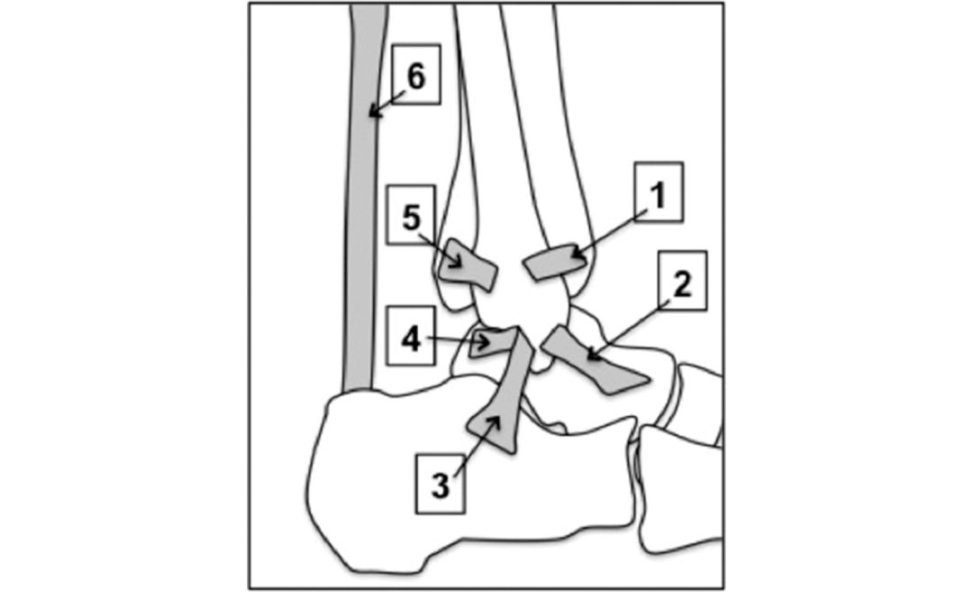
structure 3 is the calcaneofibular ligament and structure 5 is the posterior distal tibiofibular ligament.
What structures are injured in a lateral ligament injury?
The mechanism is usually a rotational injury with sequential failure of the ligaments from front to back, hence the anterior talofibular ligament or ATFL is most commonly injured followed by the calcaneofibular ligament or CF Land the posterior talofibular ligament is the least frequently injured.
How would you go about diagnosing a lateral ligament injury to the ankle?
There may be a history of recurrent sprains and instability. In apa tien t with a chronic history the clinical signs of instability would be a positive anterior drawer testor talar tilt t est.
Tell me more about those two tests.
Pain or excess anterior translation or a sulcus sign developing at the anterolateral corner of the ankle are signs of an ATFL injury. The other ankle must be examined for comparison. The lack of a firm endpoint or tilt in excess of the normal side suggests instability and the CF Lis considered to be injured if this testis positive.
What other clinical findings may be positive in apa tien t with recurrent ankle sprains?
Ankle sprains are more common inpatients with a cavus foot or hypermobility.
If you suspect a lateral ligament injury how will you proceed in managing this patient?
The first step in management would be rehabilitation with physiotherapy, concentrating on peroneal strengthening and proprioceptive training.
What percentage of patients recover?
The vast majority.
[That’s vague] Do a figure?
Sorry. Around 20% of patients develop symptoms of chronic ankle instability such as recurrent sprains, weakness and instability. Candidates should know this and may lose a mark.
How do you determine severe injuries?
The options for acute grade III injuries include cast immobilization or functional management. Cast immobilization involves 3 weeks in a below-knee walking cast followed by 12 weeks proprioceptive rehabilitation. Functional management involves early mobilization with external support and a protocol of rest, ice, compression and elevation.
What if the patient continues to have significant symptoms despite adequate rehabilitation?
Other investigations includes tress X-rays of the ankle and/or ultrasonography to assess the degree of ligamentous injury and if the patient is still having significant pain and swelling an MRI scan to look for additional pathology.
What other conditions would you be looking for?
My differential diagnosis for an ankle sprain that does not improve would be peroneal tendon pathology such as a split tear or subluxing tendons,
Do any scoring systems for chronic lateral instability?
No, sorry. Karlsson score, American orthopaedic foot and ankle score (AOFAS) and the foot and ankle outcome score (FAOS).
What are the surgical options for management of an isolated lateral ankle ligament complex injury in a young patient who has failed to respond to non-operativ e treatments?
The options f all into three broad categories. (1) Anatomic repair, (2) non-anatomic reconstruction, (3) anatomic tenodesis reconstruction.
What is an anatomic repair?
Anatomic repair involves the use of endogenous ligamentous tissue to restore the ligament. This is considered in cases when adequate tissue is present.
The Broström and the ‘modified Broström’ are the most widely used procedures for anatomic repair of the lateral ligament complex.
The complication rate is lower (fewer wounds, less risk of injury to the superficial peroneal nerve and decreased incidence of degenerative joint disease) compared to non-anatomical reconstructions with minimal effect on subtalar movement and a quicker rehab.
Failures have been atiribut ed to a variety of factors, including generalized ligamentous laxity, poor tissue quality , previous surgical repair, long-standing instability, and cavo-varus deformity.
What do you mean by a non-anatomic reconstruction (checkrein procedure)?
Non-anatomic reconstruction does not replicate the normal course and anatomy of the ATFL and CFL.
What do we mean by anatomical reconstruction?
These procedures utilize autogenous tendon gratis such as semitendinosus, gracilis or plantaris that are rerouted in such a way as to replicate the anatomic positions of the A TFL and CFL origin and insertion sites.
Are intra-articular lesions common in this group?
In one study associated intra-articular pathology amenable to arthroscopic treatment was identified in 83% of patients undergoing Brostrom repair [1].
Apa tien t asks how successful a ligament repair will be, what will you tell them?
I would expect a successful result in approximately 80% of patients.
What are the reported results of using a free hamstring graft?
The reports are good but there are some reports of weakness of knee flexion beyond 70°, particularly when both gracilis and semitendinosus tendons are harvested. This leaves the stabilizing evertor muscles intact together while reconstructing the A TFL and CF Land so may also be considered to be an anatomic repair.
The selected fixation device should be secure enough to maintain appropriate graft tension intra operatively to support healing and potentially allow for early joint motion
Thankyou.
Figure 5.1 Diagram of the lateral ankle ligaments.
Structured oral examination question 2#
Ankle arthritis
Describe the findings on this X-ray (Figure 5.2).
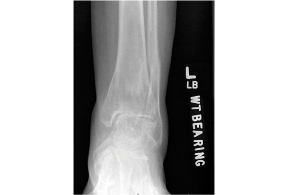
This is an AP weight-bearing radiograph of a left ankle showing narrowing of the joint space and some subchondral sclerosis.
What are the most common causes of arthritis of the ankle?
In a recent epidemiological survey, the onset of ankle osteoarthritis was atiribut able to a previous rotational fracture (37.0% of cases), recurrent sprains (14.6%), a single sprain (13.7%), pilon fracture (9.0%), tibial shaft fracture (8.5%) and osteochondral lesion of the talus (OLT) (4.7%) [2].
How is this patient likely to present?
The patient is likely to complain of pain, restriction of movement, deformity and difficulty in undertaking activities of daily living (ADLs).
Are you aware of any classification systems for arthritis of the ankle?
No, I am not aware of any classification systems specific to the ankle. The Kellgren and Lawrence Radiographic Criteria can be used [3].
The X-ray you have been shown belongs to a 42-year-old manual worker who had an ankle fracture 7 years ago that was managed non-operativ ely. Describe your management strategy for this patient.
I would first want to take a full history and examine him, then obtain a lateral standing radiograph.
Tell me about the management options a vailable for ankle arthritis.
intra-articular steroid injection or visc o-supplementation and physiotherapy. One study reported a strong positive effect on pain and function after four PRP injections a t weekly intervals [4].
What surgical options are available?
There are two types of surgical option a vailable, those aimed to ‘buy time ’ or provide temporary relief and definitive treatments. The other option is distraction arthroplasty [5]. The definitive surgical options are ankle fusion or ankle replacement.
What about arthroscopic debridement and osteophyte resection?
This may be helpful inpatients with mild arthritis with a large osteophyte restricting motion or causing painful impingement at the extremes of motion.
What about distraction arthroplasty?
Distraction arthroplasty can be used on several joints including the hip, knee and ankle to preserve the joint space and decrease the weight-bearing load by using an external fixator to distract the respective joint. Distraction arthroplasty is best suited for post-traumatic ankle osteoarthritis.
Would you offer him distraction arthroplasty?
Ankle function declines following joint distraction such that at 5 years around 50% of patients will either have gone on to ankle arthrodesis or replacement. Due to the relatively small number of studies with only level 4 evidence and no long-term follow-up I would rather offer him ankle arthrodesis [6].
Isn’t fusion an outdated treatment now that ankle replacements are available?
No, total ankle replacements are not suitable for every patient and ankle fusion is still considered the ‘gold standard’, especially so for younger patients with severe post-traumatic ankle arthritis.
which patients should be considered for ankle replacement surgery?
Contraindications include younger, more-activ epa tien ts, significant ankle instability, particularly deltoid ligament insufficiency, significant deformity, especially varus or valgus of more than 10°, peripheral vascular disease, a poor soft -tissue envelope, marked osteoporosis or avascular necrosis of the tibial plafond or talar dome.
Do anything about the types of ankle replacement available?
The earlier designs involved a two-component design such as the Agility total ankle replacement, which required fusion of the distal tibiofibular joint.
Apa tien twants to know how longan ankle replacement will last. What will you tell them?
The 10-year survival is about 85%, but there are fewer data available compared to knee and hip replacements [7–10].
The 42-year-old patient we began by discussing wants an ankle replacement. What would you tell him?
He is a young patient in a manual job. an ankle fusion would be a better option
He still w ants a replacement, as he is keen to get back to hill walking and sports and doesn’t want a stiff ankle. What will you tell him now?
He would be at risk of early failure with an ankle replacement due to his age and level of activity . Postoperative complications of total ankle replacement include infection, loosening , progressive intracomponent instability or deformity, subsidence and polyethylene failure.
What position should his ankle be fused in?
The ankle should be fused in 5° of hind-foot valgus, 10° of external rotation and the foot should be plantigrade.
What complications will you warn him about?
Infection wound healing problems,
Anything new on the horizon?
Ankle osteochondral allograft reconstruction involves replacing all or a large part of the arthritic ankle joint with a cadaveric bulk osteochondral allograft.
Thankyou.
Figure 5.2 X-ray showing ankle arthritis.
Structured oral examination question 3#
The rheumatoid foot
Please have a look at this radiographic print and tell me what you see. (See Figure 5.3.)
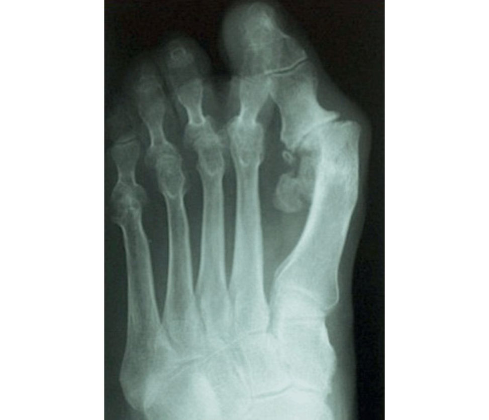
This is an AP radiograph of a forefoot. There may be deformities of the lesser t oes and I would like to see a lateral view to clarify this.
A lateral view would be very helpful. What do you think is the underlying diagnosis?
The changes suggest that this is an inflammatory polyarthropathy such as rheumatoid arthritis.
Could it be anything else?
The appearances could be secondary to a neuropathic process.
What might be the commonest neuropathic process that could cause these appearances?
A peripheral neuropathy such as that associated with diabetes mellitus would be the commonest.
How would you confirm your diagnosis?
A detailed history would be most informative Specifically, I would enquire about pain, swelling and sensory alteration.
. This lady gives a clear history of progressive, painful, bilateral small joint swelling and post-immobility stiffness. She has gr eat difficulty finding comfortable shoes and describes the feeling of walking on pebbles. She is not aware of any diabetes or sensory loss. What are your thoughts at this stage?
This appears to be an inflammatory arthropathy. This requires confirmed synovitis of one or more joints, with absence of alternative explanation for the synovitis, and achieving a score of 6 or greater out of 10 from domains including: Numbers and location of the involved joint(s). These criteria replaced the previous set from 1987, which were felt to lack sensitivity in early disease.
Yes. Her feet are making her life pretiy miserable and she would like you, as an orthopaedic surgeon, to do something to make them better. Your examination finds marked active synovitis and plantar tenderness under the metatarsal heads as well as a minimally correctable hallux valgus. There is some hammering of the lesser toes with a cock-up deformity of the second toe. Sensation and perfusion appear good. What are you going to do?
First, I would want to know if she is known to a rheumatology service and has had any attempt at non-operativ e intervention.
She has never seen a rheumatologist and has never sought help for her feet other than from you via her GP.
I would advise her that operations are helpful but that she should be formally assessed by a rheumatologist to confirm the diagnosis and achieve disease control using DMARDs. The key buzz phrase to mention (if appropriate) is that patients with rheumatoid arthritis require a (contemporary) multidisciplinary approach to their management.
I think that is appropriate advice at this stage. However, she returns to you a year later. Her synovitis is controlled by biological agents, but she has not found insoles and modified shoes helpful. How would you manage her at this point?
I would suggest surgery in the form of forefoot reconstruction.
Why?
This is a proven intervention with good results.
How good?
More than 80% of patients report significant improvement.
Would you fuse or excise the first metatarsophalangeal joint?
in a low-demand patient excision is associated with reduced complications and more rapid rehabilitation [ 11].
How would you secure the arthrodesis?
I would use an oblique compression screw augmented by a dorsal locking plate as biomechanical and clinical studies have shown this to be the most reliable method.
Would you always excise the lesser metatarsal heads in apa tien t of this age who now appears to have their disease under control?
No. It would be appropriate to perform shortening osteotomies such as Weil osteotomies to preserve the metatarsal heads if they are not badly diseased.
Surely that just prolongs the procedure and increases the risk of complication?
Yes, but it is very difficult to salvage a rheumatoid foot without metatarsal heads if the disease progresses in subsequent years.
Tell me about the principles of surgery in rheumatoid arthritis.
Surgery is indicated when symptoms and/or deformity are uncontrolled or geting worse.
What steps can a surgeon take to minimize the risk of complications?
A drug history is vital, Biological agents should be stopped in the run up to surgery and not resumed untilt here is good evidence of postoperative healing. Incisions must be planned with care, both to maintain adequate skin bridges and to ensure satisfactory wound closure if significant deformities are being corrected.
How long would you stop biological agents for?
Two weeks pre- and postoperatively [12,13].
What about other disease-modifying anft-rheuma tic drugs? Which other ones would you stop?
Peri operative management of RA medications [ 14]: Steroids: low dose (≤ 7.5 mg/day) or any dose if for < 3 weeks should be given as usual daily dose. Methotrexate: continue, as does not impair wound healing or increase peri operative infection risk.
I would like to backtrack . Would you alter your management if she also had signs and symptoms of hindfoot arthritis?
Flexible hindfoot deformity could be left un til more symptomatic.
Which hindfoot joints are most commonly affected in rheumatoid arthritis?
The talo-navicular joint is most commonly affected, followed by the subtalar and calcaneocuboid joints.
Can you outline the arguments for and against isolated talo-navicular fusion in RA?
Isolated talo-navicular fusion is a lesser procedure than triple fusion for both patient and surgeon and effectively eliminates hindfoot motion Historically, a non-union rate of up to 37% has been reported,
Thankyou.
Figure 5.3 AP radiograph of rheumatoid forefoot.
Structured oral examination question 4#
Cavus foot
These are photographs of the left foot of a 20-year old man (Figure 5.4). Describe them.
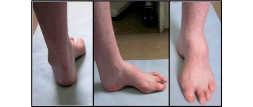
These clinical photographs show the anterior, medial and posterior views of a left foot with a cavus deformity.
What is the likely underlying cause?
The causes of a cavus foot may be broken down into congenital or acquired. The neuromuscular causes may be grouped into central nervous system disease such as cerebral palsy or Friedrich’s ataxia, spinal cord lesions such as spina bifid aor spinal dysraphism, peripheral nervous system lesions such as an HSMN or muscular causes such as muscular dystrophy. In the calcaneocavus foot the calf muscles are weak and the heel is in calcaneus and often valgus.
HSMN?
Hereditary sensorimotor neuropathies. These are a group of inherited neurological conditions. Char cot–Marie–Tooth is the most common of these conditions.
Can you go into more detail? How do these conditions lead to a cavus foot deformity?
The most commonly recognized is Charcot–Marie–Tooth (CMT) disease, which affects approximately 1 in 2500 people. Type I with an autosomal- dominant inheritance pattern tends to present in the second decade and patients have peroneal muscle weakness, abnormal (slow) nerve conduction studies, absent reflexes and hand involvement. Genetic analysi sis able to diagnose and group these conditions more accurately and at least 17 types of CM Thave been described.
What causes the deformity in CMT?
In CMT the tibialis anterior and peroneus brevis muscles are weak and the strength of the antagonistic muscles tibialis posterior and peroneus longus causes the deformity. In addition, the long extensors to the toes are recruited to assist ankle dorsiflexion, causing claw toe deformities.
What symptoms is this patient likely to complain about?
instability of the ankle with a history of frequent ankle sprains and deformity of the foot with problems fiting footwear or alteration of g ait.
What are the main findings you would look for in the examination of a cavus foot?
On general inspection I would be looking to see if the deformity was bilateral and whether there were stigmata of a generalized condition such as intrinsic muscle wasting involving the hands. I would undertake a neurological examination of the lo wer limbs to assess sensation, deep tendon reflexes and power of the major muscle groups, particularly the tibialis anterior (ankle dorsiflexion), tibialis posterior (inversion), peroneal longus (resisted plantar flexion) and peroneus brevis (eversion). I would also like to see the patient walk to see if they had a broad-based ataxic gait (Friedrich’s ataxia) or drop-foot gait.
What is shown in these diagrams (Figure 5.5)?
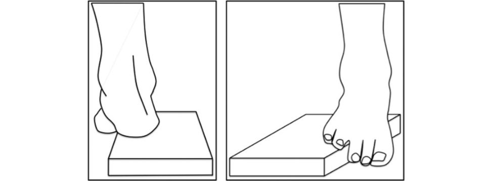
These diagrams show the Coleman block test.
And what is that?
The Coleman block test assesses flexibility of the hindfoot deformity by eliminating the deforming drive of the forefoot. If the heel assumes neutral to 5° valgus alignment when viewed from behind the hindfoot deformity is considered flexible and driven by the forefoot [15].
What investigations would you use to evaluate this foot further?
An MRI scan of the spine is required if the patient has any signs or symptoms suggesting an underlying spinal cause.
What information does the lateral X-ray provide?
Normally this lies between ±5°. This angle is normally 150° but decreases as the cavus worsens. The calcaneal pitch angle, the angle between the floor and the undersurface of the calcaneum, should be less than 30° but may be elevated in a cavus foot.
What are the principles of managing this condition?
Firstly, it is important to identify and if necessary address the underlying cause of the cavus. The patient should be assessed for neuromuscular causes and referred for a neurological opinion if appropriate. Management can be non-operativ e with the use of orthotics to try and offload pressure areas, prevent rubbing of the toes and improve stability. Surgical treatment needs to be tailored to the individual patient underlying pathology, risk of progression, level of deformity and muscular imbalance. No single surgical procedure is appropriate for all patients and frequently, multiple procedures are required.
Thankyou.
Figure 5.4 Cavus foot deformity.
Figure 5.5 Coleman block test.
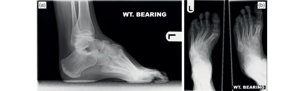
Figure 5.6a,b AP and lateral radiographs of a cavus foot.
Surgery#
1. Soft -tissue releases
Plantar fascia release
In young children, surgical release of the plantar fascia and short toe flexors may be helpful.
Gastrocnemius/Achilles lengthening
Occasionally required for contracture, but it is important to make sure a true equinus is present onstanding lateral radiographs.
2. Tendon transfers
The most common tendon transfer incavo varus foot is peroneus longus to brevis transfer to improve power of eversion. It helps stabilize the ankle.
3. Osteotomies
The osteotomy can include a closing wedge, aver tical displacement, or a combination (triplanar osteotomy).
4. Fusion
For a rigid painful foot in a young adult a triple arthrodesis can be used as a salvage stabilizing procedure that relieves pain but sacrifices joint motion.
Complications include development of ankle arthritis, pseudo-arthrosis, residue deformity, midfoot arthritis overcorrection and AVN talus.
Structured oral examination question 5#
Acquired adult flafooot
I would like you to look at this clinical photograph and tell me what you see (see Figure 5.7a).
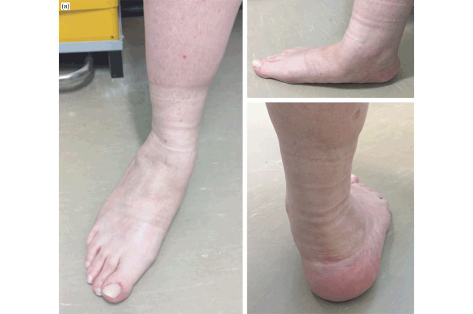
This shows the posterior view of feet in a weight-bearing stance. There is marked heel valgus, loss of the medial longitudinal arch and too many toes are visible [16].
What term is used to describe this situation?
Pes planus or flafooot.
Yes. In adults, what are the causes of this condition?
The commonest cause is tibialis posterior dysfunction. midfoot osteoarthritis and trauma (malunited calcaneum,
. How common is adult flafooot?
It is commoner in females and the incidence increases with age.
. Let’s stick with tibialis posterior dysfunction. Describe a typical patient.
The classic patient would be a female aged between 45 and 65 years with pain along the course of the tibialis posterior tendon exacerbated by activity .
What are the key examination points you would look for?
The most useful testis the ability to perform a single heel raise. I would also assess for hindfoot flexibility. These would guide classification and treatment.
What are the origins and insertions of the tibialis posterior tendon?
It arises from the posterior tibia interosseous membrane and the fibula in the proximal third of the leg and runs in the deep posterior compartment of the leg.
How do you assess the strength of the tibialis posterior tendon?
The strength of the tendon is evaluated by asking the patient to attempt to invert the foot from a plantar flexed and everted position.
How does tibialis posterior dysfunction cause a flatfoot?
The function of the posterior tibial tendon is threefold: it acts as an invertor of the subtalar joint, plantar-flexor of the ankle and an adductor of the forefoot. The inversion pull of tibialis posterior on the subtalar joint locks the transverse tarsal joint, providing a rigid lever arm with which to push off following the heel-raise phase of gait. As the function of the tibialis posterior fails, the Achilles tendon remains close to the subtalar axis, thereby reducing its function as both an inverter and a stabilizer of the subtalar joint. The static stabilizers on the medial side, in particular the spring ligament and the deltoid ligament, attenuate further.
As you have mentioned classification of tibialis posterior dysfunction could you tell me any more about this?
Johnson and Strom proposed a three-stage classification in 1989. In stage 2 disease, there is a flexible planovalgus deformity and weakness of single heel raise. In stage 3 disease, the valgus hindfoot deformity has become fixed and reconstruction is not possible. With stage 4, there is additional tilting of the talus in the ankle mortise leading to significant ankle arthritis secondary to valgus strain.
. How would you investigate this patient?
Weight-bearing AP and lateral radiographs of both the foot and ankle would help to assess structural change and exclude other causes of flafooot.
Would the arch index influence your management?
No. It is mainly used as a research tool.
Is there a place for MRI?
MRI is not routinely needed for the diagnosis of flatfeet. MRI is valuable if surgical management is being planned. It is also useful for assessing the medial structures such as the deltoid and spring ligaments and also the peroneal tendon [18].
Tell me what the treatment options are.
For stage 1, I would offer debridement of the tendon followed by 6–8 weeks of casting or splintage, then provision of a definitive arch support orthosis [19]. For stage 2 disease I would offer either a lateral column lengthening or a medializing calcaneal osteotomy in conjunction with a FDL transfer to augmentor replace the tibialis posterior [20]. In stage 3 disease, triple arthrodesis is recommended [21].
You seem very clear about surgical options Whatabout non-operativ e treatment?
I should have mentioned that. Second, it may control progressive heel valgus and flattening of the medial archIt’s not great viva tactics to jump straight in to discussing surgical options for a condition unless it is very clear that’s what the examiners want to discuss.
You spoke about an FDL transfer. Tell me about this procedure.
After obtaining informed consent I would make an incision in the line of the posterior tibial tendon, starting posterior to the medial malleolus. The free FDL tendon is then passed through a tunnel drilled in the navicular and sutured back to itself under tension.
In what direction would you pass FDL through the navicular?
From plantar to dorsal.
What is the aim of a medializing calcaneal osteotomy?
The calcaneal osteotomy directly reduces the heel valgus and brings the weight-bearing axis closer to the long axis of the leg.
When obtaining consent, what would you advise about flexion of the toes after harvesting the flexor digitorum longus?
I would expect flexion of the lesser toes to be maintained by the flexor hallucis longus via the knot of Henry.
Can you tell me alit ile more about the knot of Henry?
There are a number of fibrous interconnections between the two tendons that afford a degree of cooperation in movement.
What would you tell the patient about the success rate of the operation?
[22] reported that FDL transfer with medializing calcaneum osteotomy provided long-term pain relief and improved function in 85% of patients after a mean follow-up of 15.2 years.
Is there a place for any additional procedures such as first ray fusions or lateral column lengthening?
One concern with stage 2 adult-acquired flafooot deformity is that there is a large variability in disease severity within the classification group including hindfoot valgus, medial column stability, forefoot abduction and supination.
What approach would you use for a triple arthrodesis to correct significant, fixed valgus heel deformity?
There are advocates of triple arthrodesis via a single medial approach, but this is difficult and not always possible.
One final question. An y new developments?
PRP injections have recently been used in the early stages of posterior tendon dysfunction. We are awaiting randomized controlled data.
Thankyou.
Figure 5.7a Acquired adult flafooot. Loss of medial arch. Valgus heel, ‘too many toes sign’.
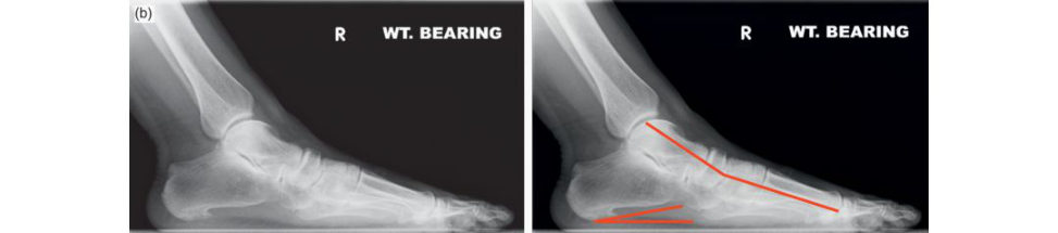
Figure 5.7b Midfoot sagift al radiograph sMeary’s angle negative (> 10°). Reduced calcaneal pitch angle (10° approx., normal 20–30°).
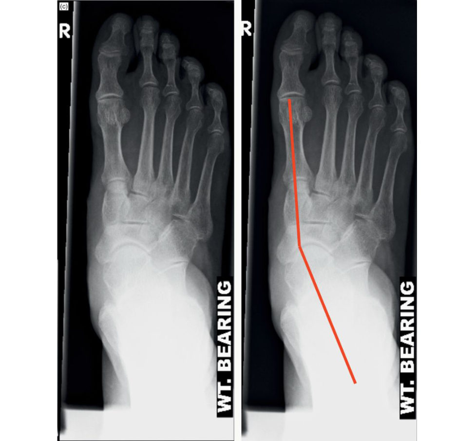
Figure 5.7c Anteroposterior (AP) radiographs of midfoot abduction. T alo to first metatarsal an angle > 10°.
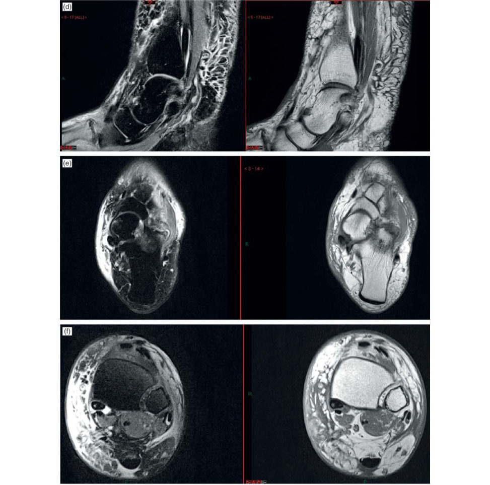
Figure 5.7d–f MRI tibialis posterior tendinopathy with fluid around tendon. Some increased signal within the tendon substance.
Structured oral examination question 6#
Hallux valgus
Please have a look at these clinical photographs and tell me what you see (see Figures 5.8 and 5.9).
These show a frontal view of a pair of feet and an oblique view of the left foot. The toenails appear friable and there is some excoriation around the lesser toes of the right foot.
. This 65-year-old lady had her left second toe removed some years ago for a presentation similar to that which she now has on the right. Her left-sided symptoms have also recurred. How would you assess her further?
I would obtain a detailed history, looking to clarify the main source of her symptoms. Can I ask what symptoms she has?
What do you think they are likely to be?
I would expect she has pain from her bunions and toes caused by rubbing on footwear and each other.
Let’s say she has all these symptoms to varying degrees. Tell me about your further assessment.
I would complete the history, including questioning about relevant conditions such as diabetes, inflammatory arthritis vascular disease and neuropathy, and proceed to examination. I would palpate for areas of tenderness, paying particular attention to the hallux MTP joint and lesser metatarsal heads. Neurovascular status will also be assessed.
You spoke about assessing the range of movement of the involved joints. Can you be more specific?
I would want to assess the range of plantar and dorsiflexion of the hallux MTP joint.
we might come back to that. Outline the value of plain radiographs in the management of hallux valgus.
I would obtain weight-bearing AP and lateral radiographs of the foot.
Why weight-bearing?
Non-weight-bearing views tend to underestimate the severity of hallux valgus, and may give misleading information about alignment and relative metatarsal length.
Keep going. What angles?
I would measure the intermetatarsal, hallux valgus and the distal metatarsal articular angles.
What is the normal range of these angles and how would these influence your management? Can you demonstrate these angles on the radiographs (Figures 5.10–5.13)?
the hallux valgus angle should be less than 15° and the distal metatarsal articular angle is normally a maximum of 15°.
What about these radiographs?
These demonstrate severe hallux valgus with an HVA 50°, IMA 22° and DMAA 8° (Figures 5.14–5.16).
If this lady had an intermetatarsal angle of 15° on the right with a hallux valgus angle of 35° and minimal passive correction of the hallux, what surgery would you plan?
If the first tarso-metatarsal joint is not lax, I would plan a scarf osteotomy combined with a lateral release and an Akin osteotomy of the proximal phalanx if necessary.
Why would you choose a scarf osteotomy?
It is a very versatile procedure with stable fixation allowing early postoperative mobilization without a cast. It can also be used to shorten or lengthen the metatarsal [23].
How would you secure the osteotomy?
With two headless compression screws.
Why not use a simpler procedure such as a chevron or Mitchell osteotomy?
For the degree of deformity described, A further disadvantage of a Mitchell osteotomy is that it produces shortening of the first metatarsal, which could lead to transfer metatarsalgia.
Can you draw a scarf osteotomy?
No, sorry.
For your proposed management, what complications would you discuss when seeking consent?
Firstly, I would advise that while early weight-bearing is possible with a scarf osteotomy, it takes up to a year for the foot to fully set ile after such surgery, but that typically 85% of patients are pleased with the outcome. I would advise a 1% risk of deep infection and as lightly higher risk of superficial infection Recurrence is possible with time, although the risk of this is greatest in adolescent cases.
How would you treat hallux varus?
A subtle varus may improve as the patient returns to normal footwear. arthrodesis of the first MTP joint arthrodesis is a reliable option in the presence of significant stiffness or arthrosis.
you have successfully treated this lady’s right foot and she is pleased with the result. Would you go ahead and do the same on the left?
No. The absence of the second toe predisposes to recurrence and I would propose arthrodesis of the hallux MTP joint.
One final question. What do we mean by a congruent versus incongruent hallux valgus (see Figures 5.17 and 5.18)?
If there is no lateral subluxation then the joint is congruent. An incongruent joint exists when the toe is in a valgus orientation and the articular surfaces do not align properly or concentrically.
Thankyou.
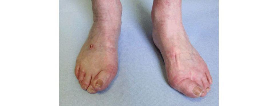
Figure 5.8 AP view of hallux valgus.
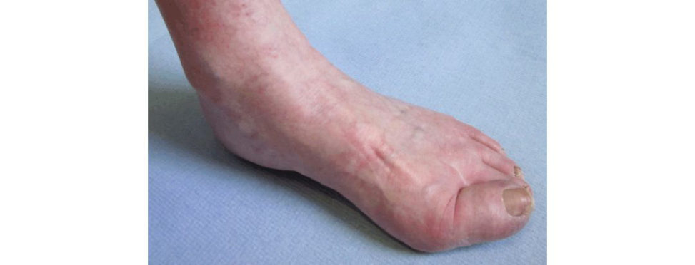
Figure 5.9 Oblique view of hallux valgus.
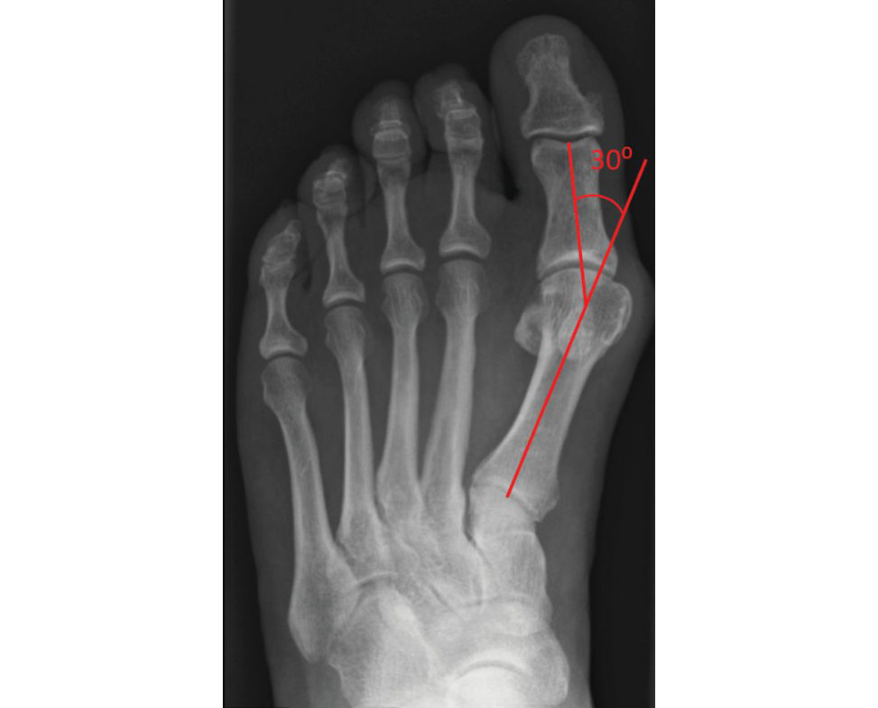
Figure 5.10 Hallux valgus angle (HVA). Angle between long axis of first metatarsal shaft and long a xis of first proximal phalanx. Normal < 15°. Mild 15–20°. Moderate 20–40°. Severe > 40°.
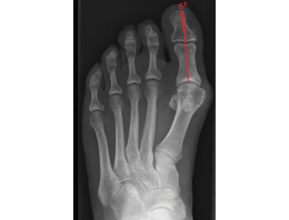
Figure 5.11 Hallux valgus interphalangeus angle (HVI). Angle between long axis of first distal phalanx and long axis of first proximal phalanx. Associated with congruent hallux valgus. Normal < 10°.
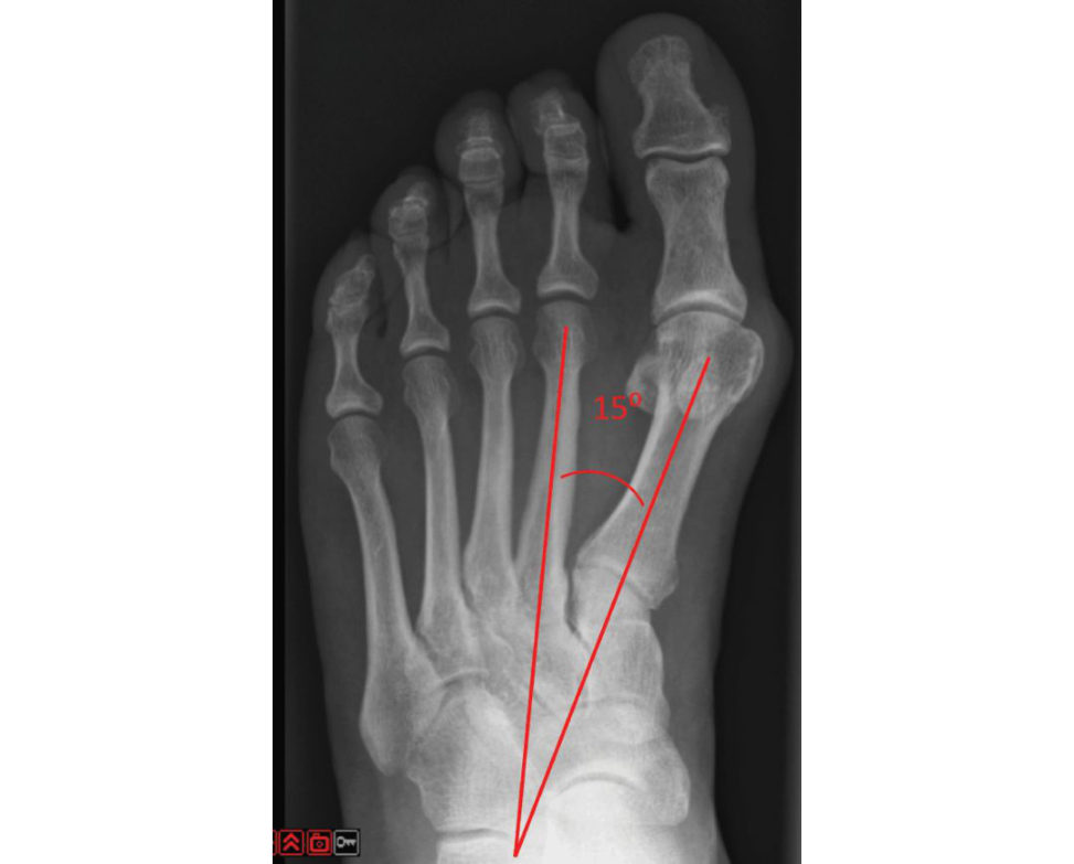
Figure 5.12 First to second intermetatarsal angle (IMA). Angle between long axis of first and second metatarsal shaft. Normal < 9°. Mild < 11°. Moderate 12–15°. Severe > 15°.
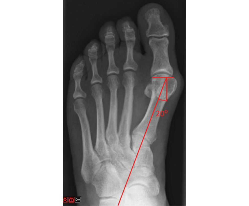
Figure 5.13 Distal metatarsal articular angle (DMAA). Angle between long axis of first metatarsal and perpendicular to a line along the articular surface. Increased angle associated with a congruent deformity. Normal < 10°.
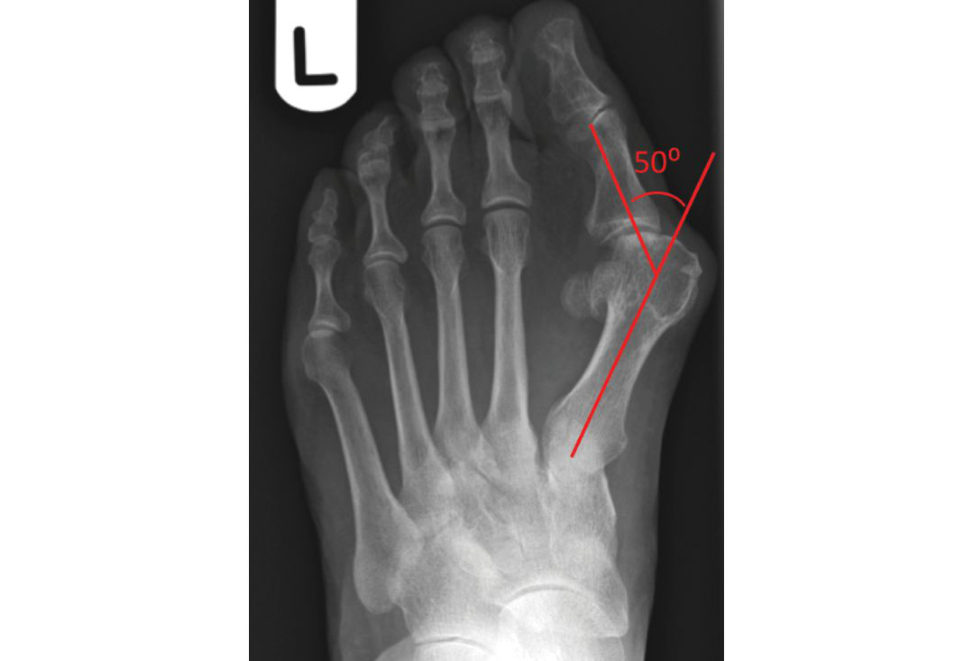
Figure 5.14 Hallux valgus angle (HVA) (pronation oft oe). Normal < 15°.
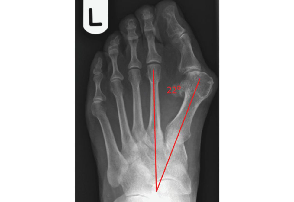
Figure 5.15 Intermetatarsal angle (IMA) 22°. Normal < 9°.
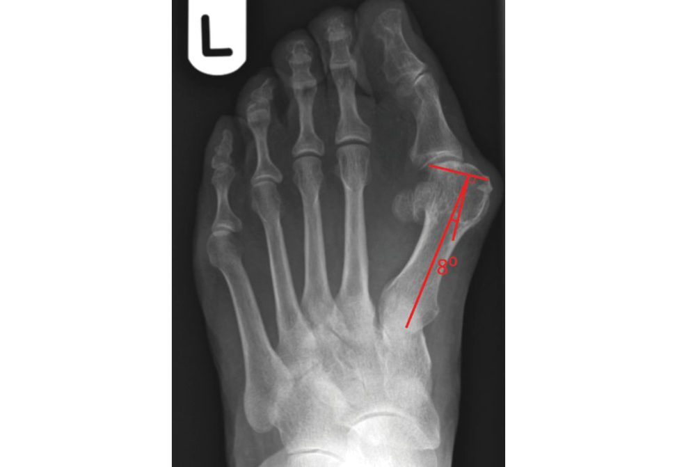
Figure 5.16 Distal metatarsal articular angle (DMAA). Normal < 10°.
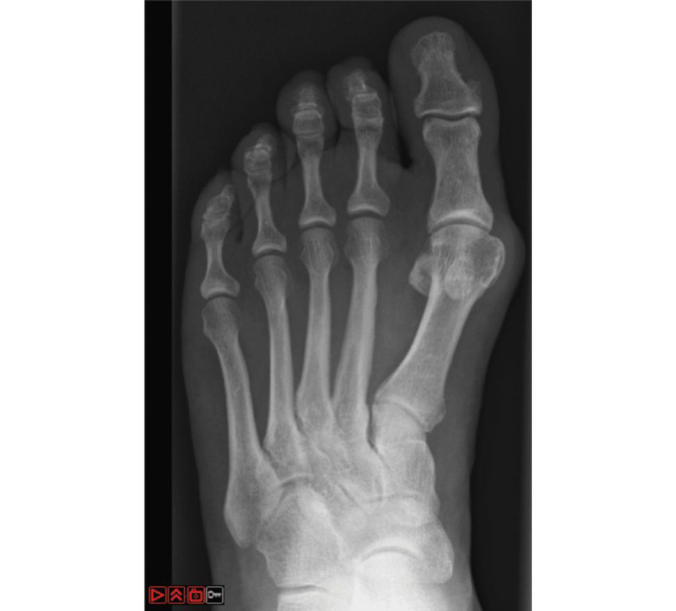
Figure 5.17 Congruent hallux valgus. Increased HVA, increased DMAA and congruent joint.
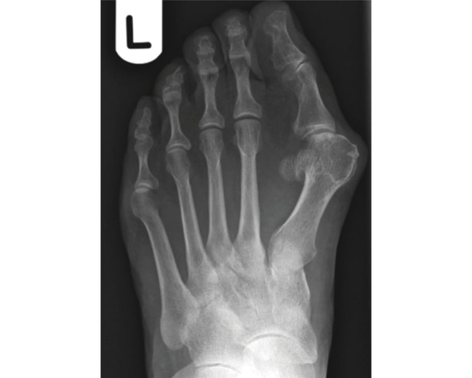
Figure 5.18 Incongruent hallux valgus. Increased HV Anormal DMAA and incongruent joint.
Structured oral examination question 7#
Hallux rigidus
This 45-year-old male patient has presented with pain and stiffness of his right big toe. Describe the X-ray findings (see Figure 5.19).
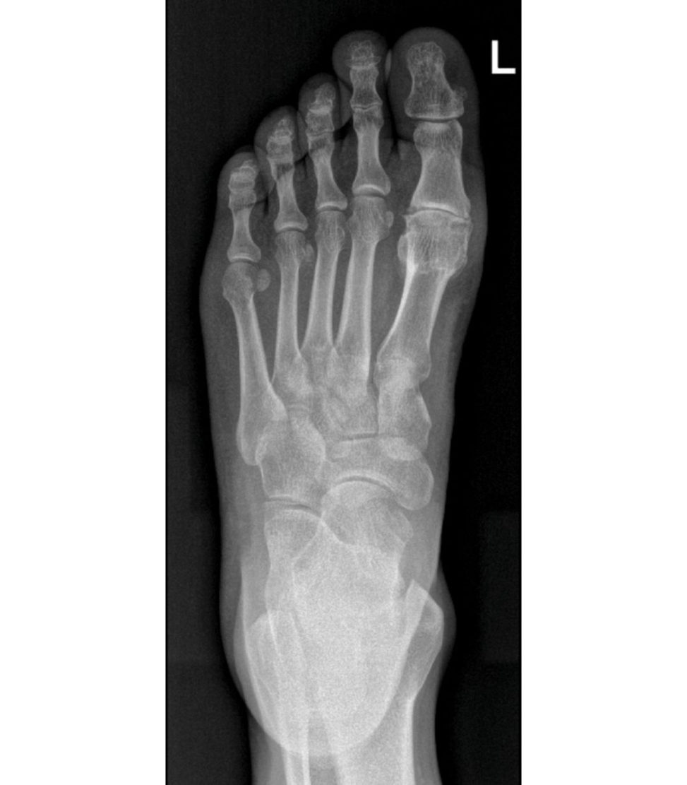
This is a radiograph of a right foot showing loss of joint space, The reis also a mild hallux valgus deformity.
what is this commonly called in orthopaedics?
Hallux rigidus.
Tell me the range of movement of a healthy first MTP joint.
The joint should be able to dorsiflex between 70 and 90° and plantarflex between 24 and 40°.
How would you manage this patient?
I would also obtain a weight- bearing lateral and an oblique X-ray of the foot in addition to the AP view we have here.
. If we concentrate on the clinical examination, what specific findings are you looking for to help with your management decision?
I would need to assess the integrity of the skin and the neurovascular status of the foot. There may be medial-sided numbness present if the medial dorsal cutaneous nerve is compressed by an osteophyte. A grind test of the joint would be informative [24]. This testis usually done with the MTPJ in relative neutral dorsiflexion and can be a pointer to articular cartilage involvement [25].
What is the importance of the IPJ?
Fusion of the first MTPJ may accelerate degeneration in the surrounding joints so if the IPJ is already symptomatic a motion-pr eserving procedure at the MTPJ may be more appropriate.
Are you aware of any grading systems for this condition?
The most widely used is a radiographic grading by Hatirup and Johnson in which Grade 1 is a well-preserved joint space with mild to moderate osteophytes,
Right, so talk me through the management options for apa tien t with hallux rigidus.
In the first instance I would advise non-operativ e treatment. The options include activity modification, NS AIDS, footwear modification or an in tra-articular steroid injection.
And the operative options?
The choice depends on the grade of the arthritis, patient symptoms and expectations.
then, back to the operative options for treatment.
In Grade 1 or 2 disease, a cheilectomy, in which the osteophytes and the dorsal 25–30% of the articular surface are resected. Satisfaction rates of 90% have been reported, with improvement in dorsiflexion from 20 to 40°. For Grade 2 or 3 disease inpatients who are young and still highly active I would combine a cheilectomy with a Moberg dorsal closing wedge osteotomy of the proximal phalanx. Arthrodesis of the first MTPJ is still the mainstay of treatment for severe disease and joint preparation with dome-shaped reamers and a lag screw and dorsal plate construct is the most biomechanically sound fixation [ 30].
back to arthrodesis. What is the optimal position for fusion?
Dorsiflexion of 25° across the MTPJ, valgus of 10–15° and neutral rotation to ensure an effective plane of motion of the IP J.
How will you consent apa tien t for arthrodesis of the first MTPJ?
I will explain that the aim of surgery is to relieve pain and optimize mobility. damage to the medial cutaneous nerve,
If we return to the patient we started discussing. He is a 45-year-old male who is a keen walker. He has significant stiffness and pain on mobilization and dorsiflexion, but a grind testis negative. He has exhausted non-operativ e measures. What treatment will you offer him?
I would offer him a cheilectomy with a proximal phalanx osteotomy as this has the added benefit of improving range of movement over a cheilectomy on its own,
He is not keen on the joint being stiffened up and has read on the internet about joint replacements and is keen for this.
I would inform him that for his age group there would be a significant risk of loosening of the prosthesis resulting in failure, thus the need for revision surgery. They reported a 20.9% rate of prosthesis loosening causing instability and pain during gait and an 11% chance of revision surgery being required.
He is still not convinced.
I would refer him to a colleague for a second opinion. [Bell] For the FRCS (Tr & Orth) exam joint replacement is very controversial.
Figure 5.19 X-ray showing hallux rigidus.
References
1. Kibler WB. Arthroscopic findings in ankle ligament reconstruction . Clin Sports Med. 1996;15(4):799–804.
2. Saltzman CL, Salamon ML, Blanchard GM, et al. … center. Iowa Orthop J. 2005;25:44–46.
3. Kellgren JH, Lawrence JS. Radiological assessment of osteoarthrosis. Ann Rheum Dis. 1957;16:494–501.
4. Repetto I, Bift B, Cerruft P, Trentini R, Felli L. … Foot Ankle Surg. 2017;56(2):362–365.
5. van Valberg AA, van Roermund PM, Marijnissen AC, et al. 1999;7:474–479.
6. Nguyen MP, Pedersen DR, Gao Y, Saltzman CL, Amendola … Joint Surg Am. 2015;97(7):590–596.
7. Wood PLR, Prem H, Suf ton C. Total ankle replacement: medium term results in 200 Scandinavian total ankle replacements. J Bone Joint Surg Br. 2008;90B(5):605–609.
8. Bonnin M, Gaudot F, Laurent J-R, et al TheS alto total ankle arthroplasty: survivorship and analysis of failures at 7 to 11 years. Clin Orthop Relat Res. 2011;469:225–236.
9. Mann JA, Mann RA, Hort onE. STAR ankle: long-term results. Foot Ankle Int. 2011;32(5):473–484.
10. Labek G, Klaus H, Schlichtherle R, et al. … Ankle Int. 2011;32 (8):740–745.
11. Rosenbaum D, Tim taB, Schmiegel A, et al. First ray resection arthroplasty versus arthrodesis in the treatment of the rheumatoid foot. Foot Ankle Int. 2011;32(6):589–594.
12. Lee MA, Mason LW, Dodd sAL. The peri operative use of … Orthopedics. 2010;33(4):257–262.
13. Howe CR, Gardner GC, Kadel NJ. Peri operative medication management for the patient with rheumatoid arthritis . J Am Acad Orthop Surg. 2006;14:544–551.
14. Scanzello CR, Figgie MP, Nestor BJ, Goodman SM. Peri operative management of medications used in the treatment of rheumatoid arthritis . HSS J. 2006;2(2):141–147.
15. Cole manS, Chestnut W. A simple test for hindfoot flexibility in the cavovarus foot. Clin Orthop Relat
Res. 1977;123:60–62.
16. A ‘too many toes sign’ will be present where more than one to two toes are seen along the lateral aspect of the affected foot.
17. Myerson MS, Corrigan J. Treatment of posterior tibial tendon dysfunction with flexor digitorum longus tendon transfer and calcaneal osteotomy. Orthopedics. 1996;19:383–388.
18. Abousayed MM, Alley MC, Shakked R, Rosenbaum AJ. Adult-acquired flafooot deformity: etiology , diagnosis, and management. JBJS Rev. 2017;5(8):e7.
19. Teasdall RD, Johnson KA. Surgical treatment of stage I posterior tibial tendon dysfunction . Foot Ankle
Int. 1994;15(12):646–648.
20. Myerson MS, Bade kasAS chon LC. Treatment of stage … Foot Ankle Int. 2004;25(7):445–450.
21. Kell yIP, Easley ME. Treatment of stage 3 adult acquired flafooot. Foot Ankle Clin. 2001;6:153–166.
22. Chadwick C, Whitehouse SL, Saxby TS. Long-term … . Bone Joint J. 2015;97(3):346–352.
23. Barouk LS, Toullec ET. Use of scarf osteotomy of the first metatarsal to correct hallux valgus deformity.
Tech Foot Ankle Surg. 2003;2(1):27–34.
24. This is done by rotating and axially loading the hallux MTPJ with the toe in relative neutral dorsiflexion and can be a pointer to articular cartilage involvement.
25. Patients who experience grind test pain with the MTPJ in neutral or plantar flexion often have more extensive disease than realized.
26. Hatirup SJ, Johnson KA. Subjective results of hallux rigidus following treatment with cheilectomy. Clin
Orthop Relat Res. 1988;226:182–191.
27. Moberg E. A simple operation for hallux rigidus. Clin Orthop Relat Res. 1979;142:55–56.
28. Taranow WS, Moutsatson MJ, Cooper JM. Contemporary … Foot Ankle Clin N Am. 2005;10:713–728.
29. Carpenter B, Smith J, Motley T, et al. Surgical treatment of hallux rigidus using a metatarsal head resurfacing implant: mid-term follow-up. J Foot Ankle Surg. 2010;49:321–325.
30. Polift J, Hayes J, Njus G, et al. First metatarsal–phalangeal joint arthrodesis: a biomechanical assessment of stability. Foot Ankle Int. 2003;24(4):332–337.