Chapter 6 Spine
Prasad Karpe
Introduction#
but can pop up in basic science (structure of intervertebral disc), trauma (thoracolumbar fractures and their management) or paediatrics (adolescent scoliosis).
Also, spine is like maths –neurology and level of pathology should add up. Besides, indications for surgery are specific and usually encompass neurology or instability.
Each question is to be answered in 5 minutes. This needs a lot of planning and practice.
By this time, you will be well aware of the marking process: minimum 6 pass for each question.
7 or 8 at most stations . Try to stay calm and be positive And no matter how bad the previous question was, forget it and recompose for the next question.
The usual format for a viva would be that a candidate would be shown a radiological picture (X-ray/CT/MRI) or a clinical picture and asked to describe it.
Describing an X-ray#
Describe what X-ray it is first
PA and lateral standing X-ray of lumbar/thoracic/cervical/full spine showing ...
Mention immature skeleton if you see a physis.
Lateral X-ray
1. Normal curves – cervical and lumbar lordosis, thoracic kyphosis.
2. Abnormal curves – exaggerated thoracic kyphosis, loss of lumbar/cervical lordosis, cervical kyphosis.
3. Disc height maintained or lost at any level. Fuzzy endplates could be a sign of discitis.
4. Facet arthritis.
5. Primary canal stenosis as evident by short pedicles.
6. Fracture – compression, burst, stable or unstable fracture.
7. Sagift al balance of full spine weight-bearing lateral – normal, positive or negative.
8. Listhesis – is it isthmic (if lysis of pedicles seen) or degenerative facet arthropathy)?
Meyerding grading.
Pelvic tilt, sacral slope and pelvic incidence.
AP view
1. Cancer – winking owl sign – destruction of pedicle duet o cancer.
2. Degenerative – facet arthritis or scoliosis (curve can be in either direction).
3. Idiopathic scoliosis – usually right convex in the thoracic spine (away from the heart).
4. Prolapsed disc – list away from nerve compression.
5. Evaluate pedicles always on AP view. Absent pedicle may beseen in metastases, aneurysmal bone cyst, osteoblastoma, trauma or congenital absence of pedicle.
Detecting the correct level
1. Highest point on the iliac crest usually points to L4/5 on the lateral view.
2. Count from C2 downwards if you have a full spine X-ray.
3. The tweltih rib can be helpful.
If confusion still exists between sacralization of L5 and lumbarization of S1, then it would be safe to comment on pathology based on the last mobile level.
History and examination#
By the time you have described the radiograph, you know what the clinical scenario is and so a focused history is not a difficult task.
Every clinical scenario has some important questions that need to be asked. Examples include:
Sciatica – it is necessary to know the exact location of the leg pain (dermatome) as this will clinch the nerveroot on the history alone.
Night pain – cancerin elderly.
Back pain worse than leg pain or significant back pain could mean instability.
An immunocompromised patient such as IVDU or a diabetic gives a clue of infection.
Please read the spine clinical cases chapter in the Postgraduate Orthopaedics book for the relevant questions in his tory-taking and examination.
As for clinical examination, always divide into general and local/spine examination. So, points like
EW Sin sepsis or chest expansion in ankylosing spondylitis in the general examination will not be missed.
Specific points of importance in the clinical examination are:
1. PR and perianal sensations in caudae quina.
2. Reflexes is a must It differentiates upper motor neuron lesion from a lower motor neuron.
3. Pulsations in feet to differentiate vascular pathology.
4. Location of tenderness – lumbar facet, sacroiliac joint, hip (groin).
Relevant investigations and management are discussed in each scenario.
Contents#
1. Infection.
2. Spine metastases.
3. Benign tumour.
4. Cervical spondylotic myelopathy.
5. Lumbar prolapse disc with caudae quina.
6. Spondylolisthesis.
7. Ankylosing spondylitis.
8. Idiopathic scoliosis.
9. Non-idiopathic scoliosis.
10. Cervical disc prolapse.
Structured oral examination question 1#
Infection (discitis)
You are on call and you are asked to see a 45-year-old diabetic patient on the medical wards with back pain. The medics are concerned as she has raised blood markers and they can’t find any septic focus from their end.
1. What is the onset, duration and progress of her back pain? 2. What’s the exact location? 3. Is she able to mobilize? (Non-ambulatory usually suggests sinister pathology or advanced disease or instability.)
What next?
4. More details of her diabetes, is it Type 1 or 2, how well has it been controlled, is she diet- controlled/oral medications or insulin? 5. Any other septic focus including chest, urine, abdomen?
She is a Type 2 diabetic now poorly controlled, on insulin. The medics have ruled out all other septic focus. Nothing else significant on history and she is bedridden. She is struggling to sit or stand. No history of trauma.
I will then do a general and spine examination. What’s he rEWS?
This patient has no fever but a low temperature of 35 degrees. She also appears to be confused. What are you concerned about?
Sepsis is my major concern in this patient Sepsis is SIRS with documented infection.
(interrupting): What is SIRS?
SIRS is systemic inflammatory response syndrome and characterized by two or more of the following: Fever (> 38°C) or hypothermia (< 36°C). Tachycardia (> 90).
Tachypnea (> 20).
WBC >12 or < 4.
Altered mental state.
Blood glucose > 6.6 in absence of diabetes.
This patient already has two criteria needed for SIRS, namely altered mental state and hypothermia. My concerns are that she may progress to septic shock.
She also has low blood pressure and tachycardia.
I will do a detailed spine examination looking for specific point tenderness and progress to neurological examination.
She has significant tenderness in her lower lumbar spine. No sensory–motor deficit in any of her limbs. PR normal. What next?
Point tenderness in the back is suggestive of discitis. What are her reflexes like?
Does it matter?
Brisk reflexes could suggest upper motor neuron lesion.
Reflexes are normal.
I will look a t her bloods FBC, U/E, RFT, serum lactate. The trend is more important in WBS, ES Rand CRP than actual values. I will then follow the Sepsis 6 pathway. If feasible, I will ask for an MR Iof her lumbar spine which is the primary diagnostic modality for spine infections.
Please read this X-ray (Figure 6.1a,b).
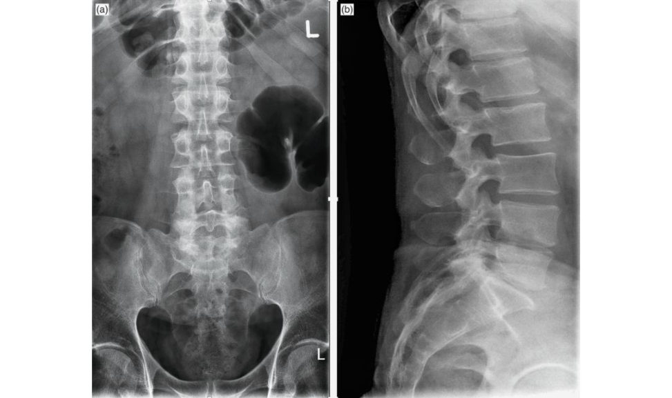
Figure 6.1a,b Anteroposterior(AP) and lateral views of the lumbar spine.
These are AP and lateral views of the lumbar spine showing destruction of the vertebral endplates at L4/5 on the lateral view.
These are her MRI scans (Figure 6.1c–g).
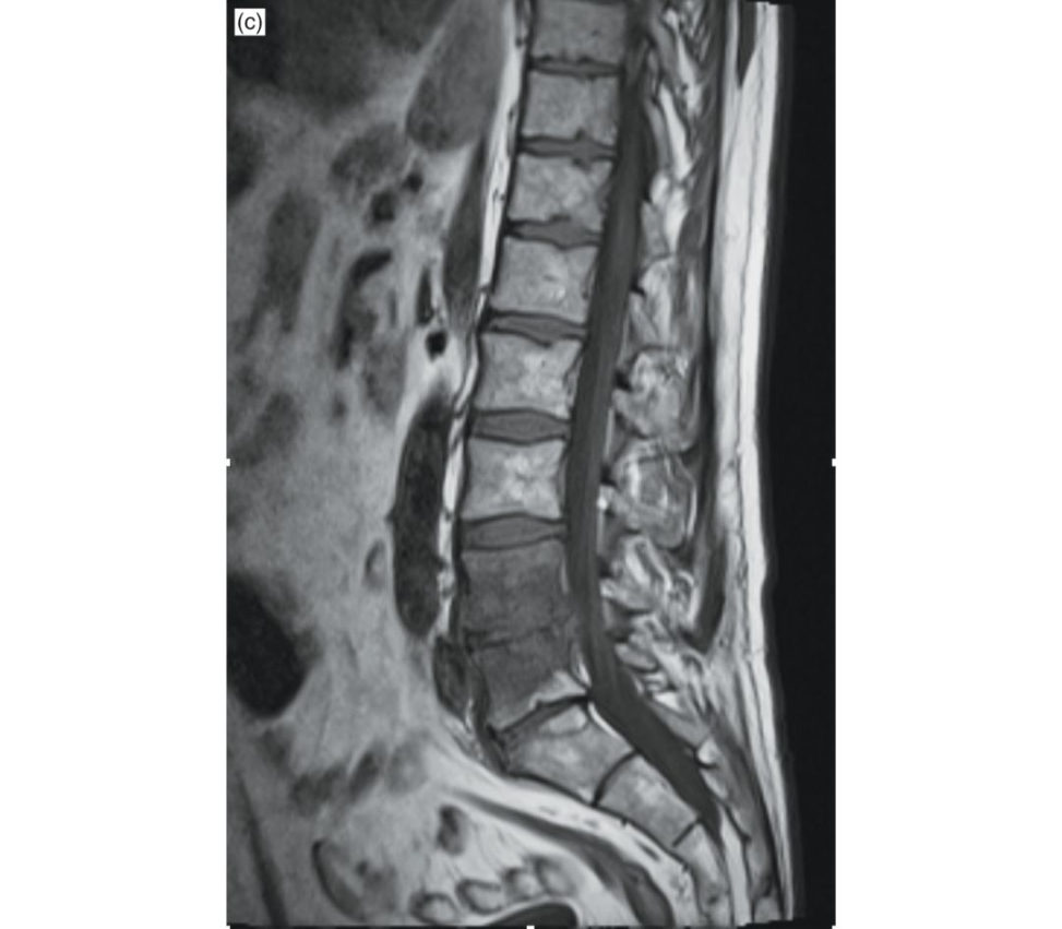
Figure 6.1c MRI lumbar spine (T1 sagift al).
This is an MRI scan showing a hypointense signal at L4/5 on sagift al T1 (Figure 6.1c) and hyperintense on sagift al T2 (Figure 6.1d). There is hyperintense vertebral marrow signal of L4 and L5 on fat-saturated T2 WI or STIR (Figure 6.1e).
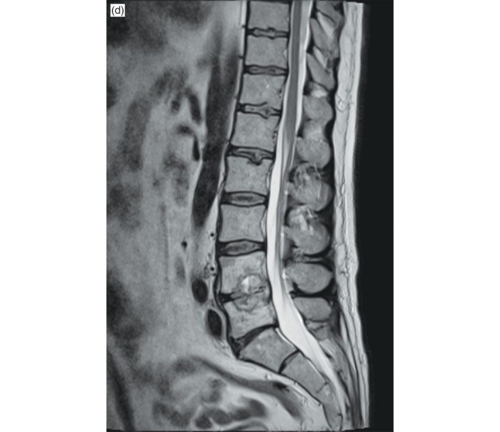
Figure 6.1d MRI lumbar spine (T2 sagift al).
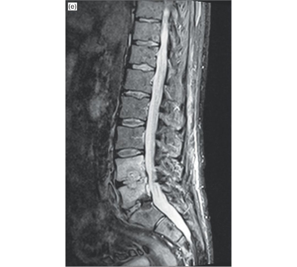
Figure 6.1e MRI lumbar spine (STIR) image.
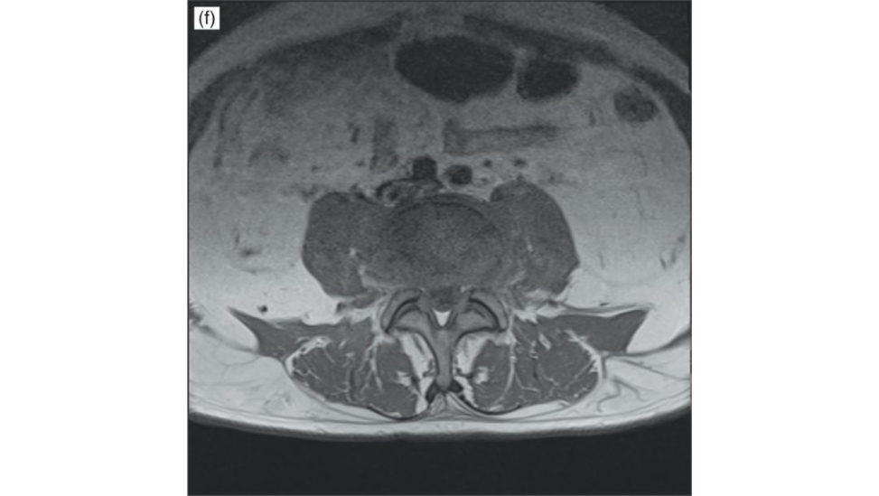
Figure 6.1f MRI lumbar spine axial cut.
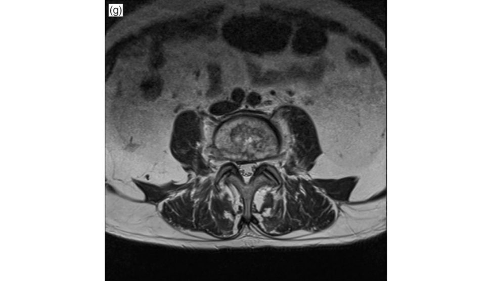
Figure 6.1g MRI lumbar spine axial cut.
Could this be metastases?
Although metastases can lead to hypointense T1 and hyperintense T2 vertebral lesions, the disc space is always spared in malignancy.
What will you do next?
The management of this patient will need a multidisciplinary approach. Treatment at this point of time is essentially supportive with the Sepsis 6 pathway. Parenteral antibiotics for 6–8 weeks in addition to a spinal brace for 6–12 weeks. Ideally, a biopsy would be ideal to decide the antibiotic. This can be done CT-guided depending on how stable the patient is. Also, a blood culture may provide a clue on the causative organism that should be done when the patient spikes a temperature.
What is the role of surgery?
2. Abscess with poor vitals to reduce the infective load, if fit for surgery. 3. Open biopsy in some cases to find exact organism and check drug sensitivity (eg. Rarely done as CT-guided biopsy has replaced it. 4. Instability – healed infection with instability.
What’s the most common pathogen? What’s the mode of spread?
Like most orthopaedic infections, Staphylococcus is the most common organism. Bacteraemia (haematogenous – arterial) from an extraspinal primary source is the most common route of infection. In children, however, the disc space infection is primary owing to vascular channels across the growth plate.
How do you monitor response to therapy?
Symptomatic improvement like generalized feeling of well-being, reduction of back pain, increased mobility. Blood parameters like ESR, CR Pand WBC. Also, serial MRI can be helpful.
This MRI is done 5 weeks after therapy. Please comment.
The hypointense marrow signal seems to be improving on the T1 image (Figure 6.1h).
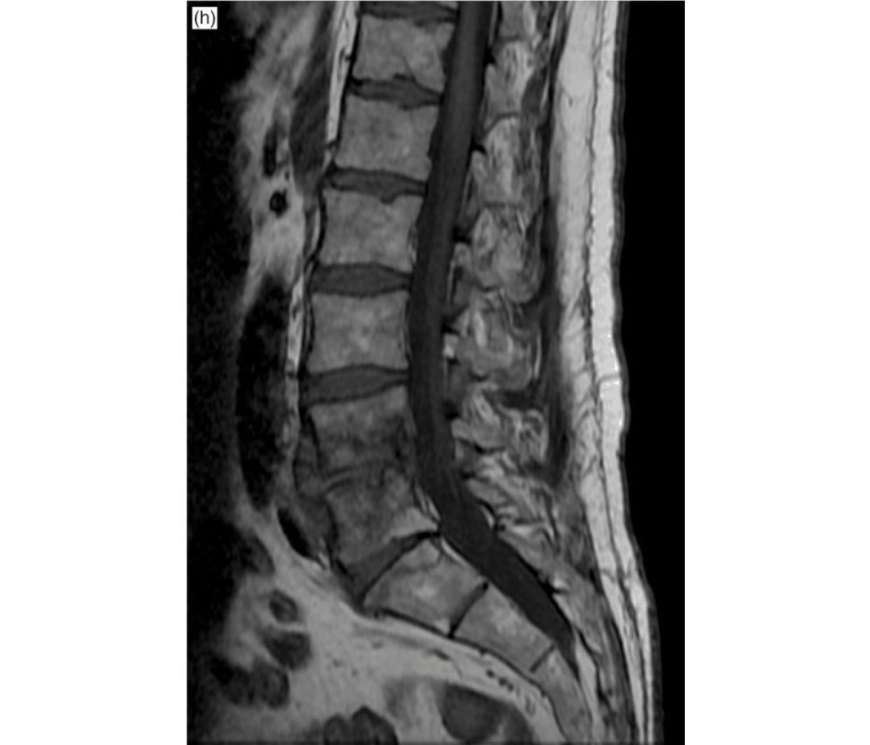
Figure 6.1h MRI lumbar spine.
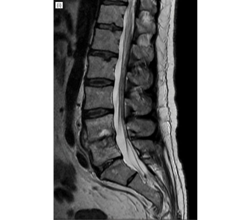
Figure 6.1i MRI lumbar spine.
Thankyou.
Learning points
1. Answer in a logical sequence of history,
2. An immunocompromised patient with either fever or joint/back pain = BONE/JOINT
INFECTION.
3. Fever is variable and can be present inless than 50% of cases. Some patients may not have an inflammatory response and fever may be absent.
4. Indications of surgery in infective spine usually are:
4.1. Neurology – worsening or advanced.
4.2. Instability (read White and Punjabi definition for instability).
4.3. Biopsy – if no improvement with antibiotics to find the causative organism, drug sensitivity or alternative pathology such as tumour.
4.4. Abscess – if no improvement with antibiotics and worsening of systemic features or neurology due to compression of neural structures by the abscess.
5. The above scenario tests management of a septic patient as well as spinal infection.
6. Adult versus paediatric spinal infection differences: vascular channels across the growth plate in children leading to primary infection of the disc.
7. Similar infective scenarios could be vertebral osteomyelitis, epidural abscess, spinal cord abscess, septic facet joint arthritis or paras pinal abscess.
Structured oral examination question 2#
Metastases
You see a 68-year-old man in your clinic with progressive back pain that’s worse at night and significant weight loss. How would you approach this patient?
His age, nocturnal pain and weight loss is suggestive of a sinister pathology such as infection or cancer. I will take a detailed history and examine ... [being interrupted]
What else do you want to know with history?
2. Neurology including bowel/bladder or gait involvement. 4. For infections – any immunocompromised state such as being on steroids, diabetes or IVDU. 5. Any other significant past history including medications, radiation or previous cancer? 6. Symptoms of other systems such as cough, abdominal pain and prostate symptoms to suggest any primary?
He is a chronic smoker with chronic cough and no other significant past medical history. He does complain of gait disturbances, though. His back pain is throughout thoracic and lumbar spine that is 8/10 on the VAS scale. There is no history of trauma. Nothing else is relevant on history.
I will proceed towards general and spine examination ... [being interrupted]
Nothing specific on general examination. Spine examination reveals tenderness along the entire thoracic and lumbar spine. He has no neurology in upper limbs but grade 3 power in lower limbs. PR is normal. Sensations are grossly normal.
What are the reflexes like?
Brisk only in the lower limbs with upgoing plantar.
possible neoplastic aetiology with chronic smoking history and significant weight loss.
Infective – FBC, CRP, ESR.
Metabolic – Ca, PO4, Alk PO4.
Neoplastic – Se electrophoresis, Se PSA, CEA.
Radiological
X-rays – full spine and chest in clinic.
MRI full spine.
These are his X-rays of lumbar spine (Figures 6.2a, 6.2b).
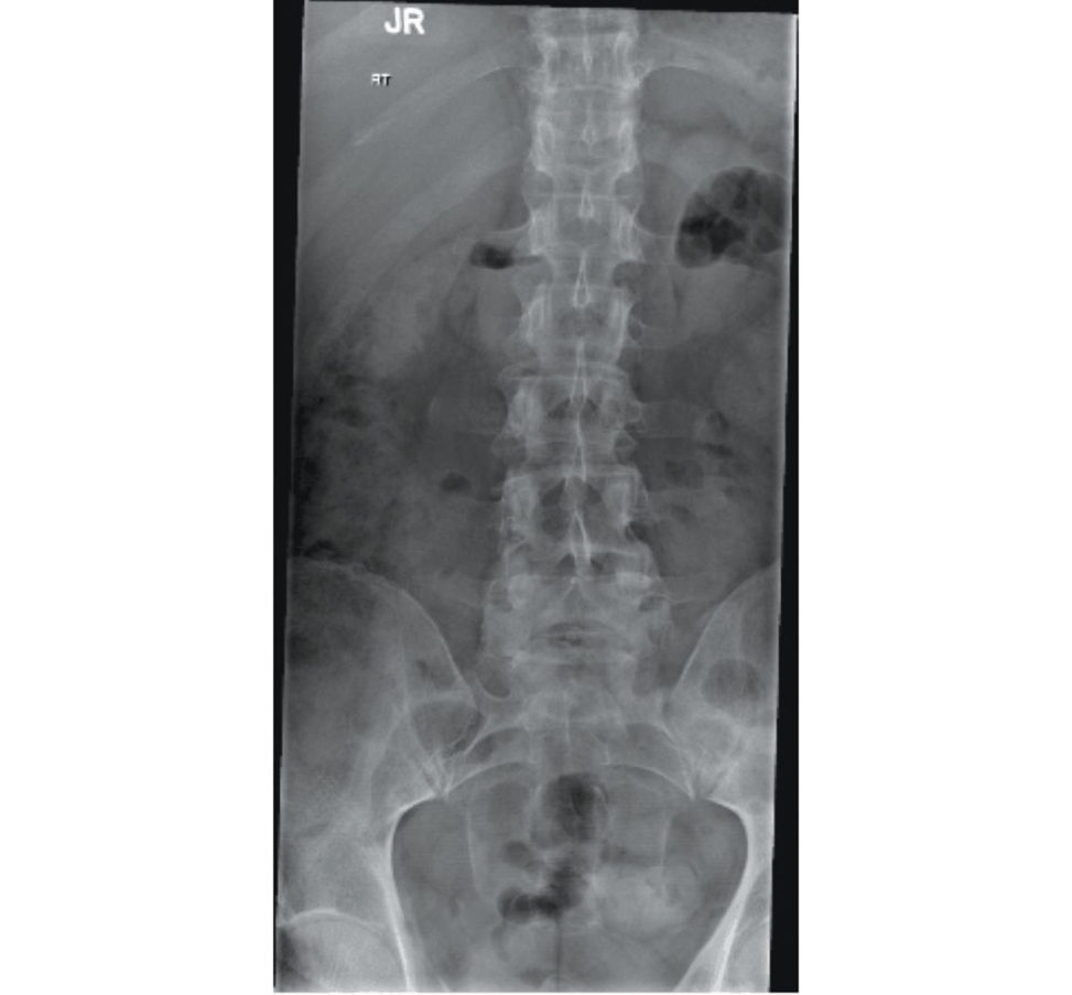
Figure 6.2a Anteroposterior (AP) radiograph of lumbar spine.
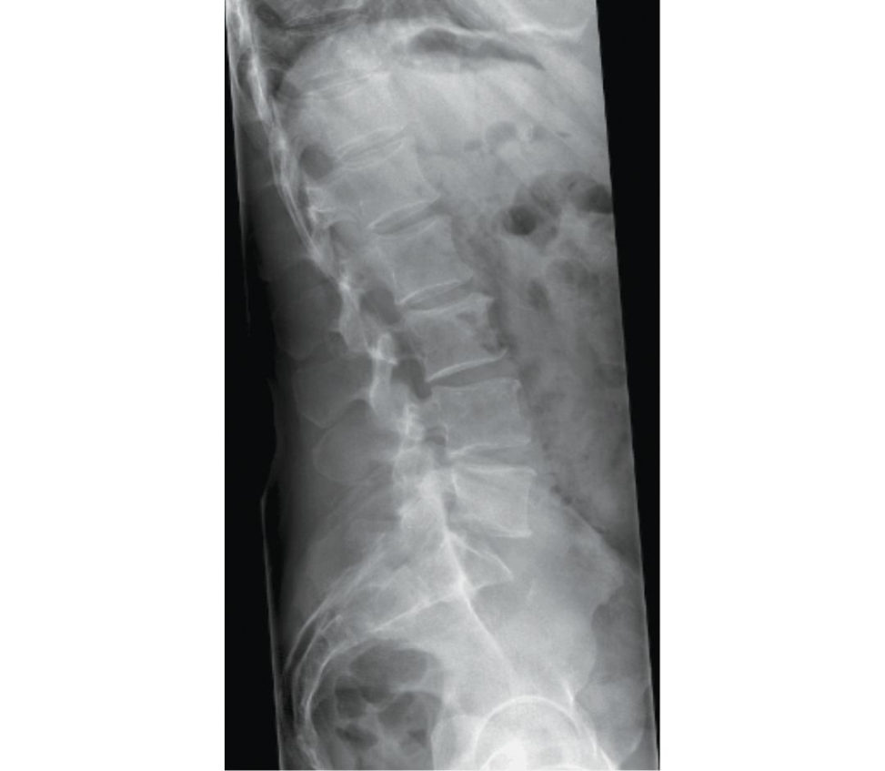
Figure 6.2b Lateral radiograph of lumbar spine.
AP (Figure 6.2a) and lateral view (Figure 6.2b) of the lumbar spine.
T11 is the only abnormality on X-rays. What next?
Because this patient has neurology, I will get him admift ed and get an urgent MRI within 24 hours.
This is his CT chest (Figure 6.2c).
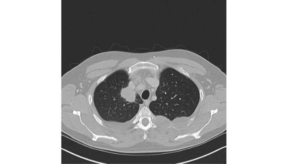
Figure 6.2c CT of chest.
suggesting a primary in the lungI will now manage this according to NICE guidelines for metastatic c ord compression 2014 [1].
Please comment on the MRI scan (Figure 6.2d).
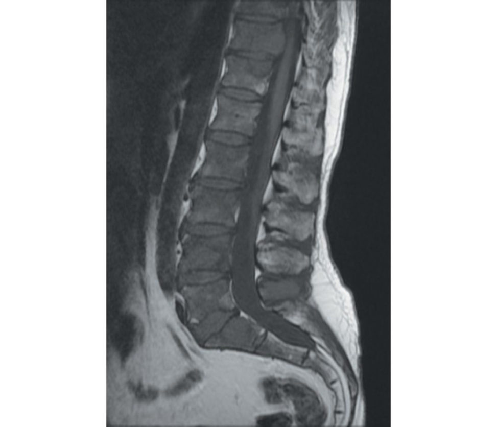
Figure 6.2d MRI lumbar spine, T1-weighted images.
MRI T1-weighted images of lumbar spine (Figure 6.2d) showing loss of normal signal of the marrow with a replacement of hypointense signal with involvement of multiple vertebral bodies.
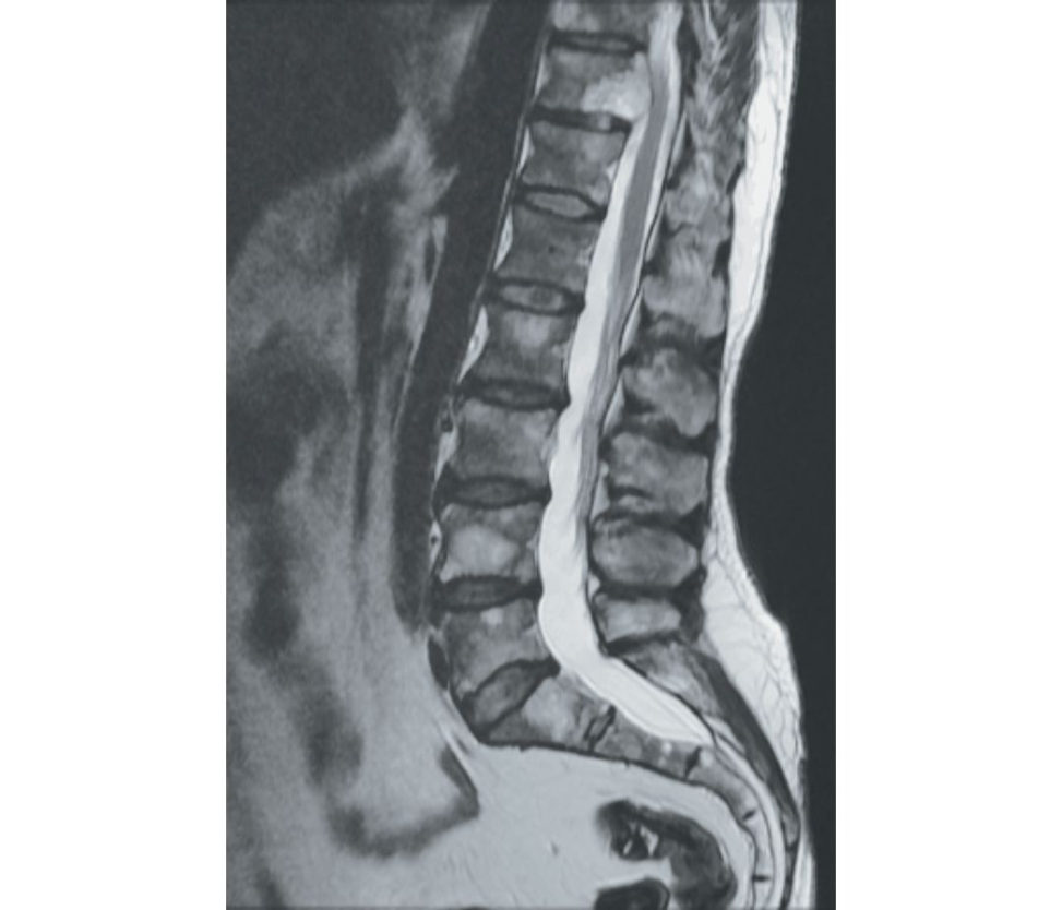
Figure 6.2e MRI lumbar spine, T2-weighted images.
Cervical MRI is normal. This is the thoracic MRI (Figure 6.2f and Figure 6.2g).
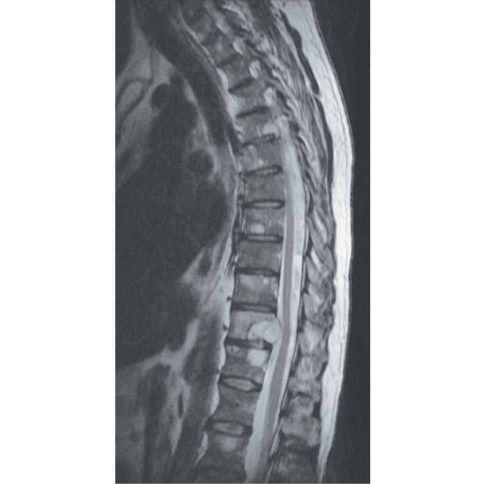
Figure 6.2f MRI lumbar spine.
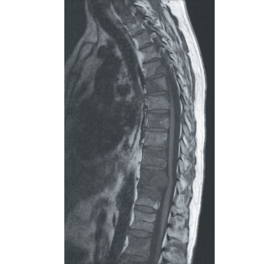
Figure 6.2g MRI lumbar spine.
Thoracic MRI sagift al images (Figure 6.2f and Figure 6.2g) show multiple-le vel marrow infiltration with the cancer.
Yes, T11 is the major level of compression. What next?
As previously mentioned, this needs to be managed according to NICE guidelines for metastatic c ord compression 2014.
Any further investigations?
I need to grade and stage this tumour.
Systemic staging is done by bone scan, CT of chest/abdomen/pelvis, PET scan or full-body MRI and is done to detect the primary and seek distant metastases.
Grading, on the other hand, is histology and is done by biopsy. high-grade undifferentiated Following the general principles applicable to all musculoskeletal tumours this biopsy should be done within the unit that will treat the tumour
Care must betaken in some metastases such as renal metastases that are very vascular and may need preoperative embolization prior to biopsy.
How would you decide about subsequent treatment?
The scoring system proposed by Tokuhashi [2] is useful in establishing indications for treatment and subsequent surgical goal. A score of less than 5 indicates a life expectancy under 1 year and a palliative approach is suggested.
His general condition is good otherwise and spine is the only metastases. What next?
Treatment options include: Surgical decompression and stabilization. Radiotherapy is mainly for palliative treatment and reduces bulk. Radiotherapy is more reserved for prostate, most breast and lymphoreticular tumours. There should be an interval of 6 weeks between radiotherapy and surgery to avoid wound problems. Here, I am more inclined for surgical instrumented decompression ± fusion depending on his fitness for anaesthesia and major surgery. Simple laminectomy to ‘decompress’ the tumour is rarely indicated. This is because the presence of the tumour most frequently found in the vertebral body is likely to lead to mechanical instability and thus kyphosis. Instrumented stabilization is frequently undertaken.
In general terms, if a curative resection is hoped for, or survival is likely to extend beyond 6 months, intervertebral bony fusion should be undertaken to avoid instrumentation failure.
How do metastases spread to the spine?
The spinal column is the most common site of osseous metastases of which thoracic spine is the most common. Spread can occur via: Direct extension – lung cancer extending into the chest wall into vertebral bodies and posterior elements.
Why do spine metastases invertebral bodies show reduced signal on T1?
Vetebral bodies contain bone marrow that has fat. This fat shows as a bright signal on T1. In metastases, this fat is replaced by cancer cells, leading to a reduced signal.
Thankyou.
Learning points
1. In cancers, the first aim is to confirm the diagnosis, then extent of spread and finally management.
2. Ask relevant questions to examiners when taking a history or asking about the findings of examination. This is just like an everyday clinic scenario.
3. Unless you ask for a relevant investigation, the examiner may not even show you the X-ray or MRI images.
4. Tokuhashi staging:
General condition (poor 0, moderate 1, good 2).
Number of extra-spinal metastases (three or more scores 0, one or two scores 1, zero scores 2).
Number of spinal bony metastases (three or more scores 0, two scores 1, one scores
2).
Number of metastases to major internal organs (not removable 0, removable 1, no mets 2).
Tissue of origin (lung/stomach 0, kidney/liver/uterus 1, other/breast/thyroid/prostate/rectum 2).
Spinal cord palsy (complete 0, incomplete 1, none 2).
Excisional surgery > 9.
Palliative treatment < 5.
5. Neurology generally points to surgery unless in a very unfit patient or a very radiosensitive tumour such as lymphoma/myeloma, where radiotherapy is recommended.
6. Myeloma – CRAB (HyperCalcaemia, Renal failure, Anaemia, Bone lesions).
7. Prognosis – median survival inpatients with metastatic bone disease.
a. thyroid: 48 months.
b. prostate: 40 months.
c. breast: 24 months.
d. kidney: variable depending on medical condition but may be as short as 6 months.
e. lung: 6 months.
8. Types of metastases:
Blastic – bone production exceeds bone destruction.
- Prostate in adults. ·
- Medulloblastoma, neuroblastoma, Ewing’s in children. ·
Lytic – bone destruction exceeds bone production.
- Renal, lung, breast and thyroid. ·
9. Differential diagnosis.
Blastic – haemangioma, Paget’s disease, osteosarcoma, benign osteoporotic compression fractures, renal osteodystrophy.
Lytic – multiple myeloma, lymphoma, spondylodiscitis (destruction of disc space with abnormal signal on either side of disc space).
10. Factors to be considered in offering treatment.
Neurology.
Type of cancer – radio-resistant, -sensitive.
Systemic disease – limited or extensive.
Instability.
References#
1. NICE. Metastatic spinal c ord compression in adults. Quality Standard 56. February 2014.
2. Tokuhashi Y, Matsuzaki H, Toriyama S, Kawano H, Ohs akaS. Scoring system for the preoperative evaluation of metastatic spine tumor prognosis. Spine. 1990;15(11):1110–1113.
Structured oral examination question 3#
Benign lesion
You see a 35-year-old fit and healthy man complaining of insidious onset LBP, more severe at night. There is no history of trauma and no red flags on history.
Is the pain localized to one specific point in his back? Any neurology?
Pain is localized to the right side of his back, for 6 months now. No neurology with no bowel or bladder symptoms. Nothing else is significant on history.
Night pain is a concern for me. If there is no other relevant history, I would like to go ahead with a general and spine examination.
Nothing remarkable on general examination. Spine examination shows right-sided point tenderness in the L45 region with normal neurology. Other joints and distal pulsations are normal.
Blood Infective – FBC, CRP, ESR. Metabolic – Ca, PO4, Alk PO4. Radiological X-rays and MRI full spine.
Why X-rays?
Night pain is a red flag. I need X-rays.
X-ray of the full spine is normal. All blood investigations are within normal limits.
I will proceed for MRI.
This is a midsagift al T2 image (Figure 6.3a). Please comment.
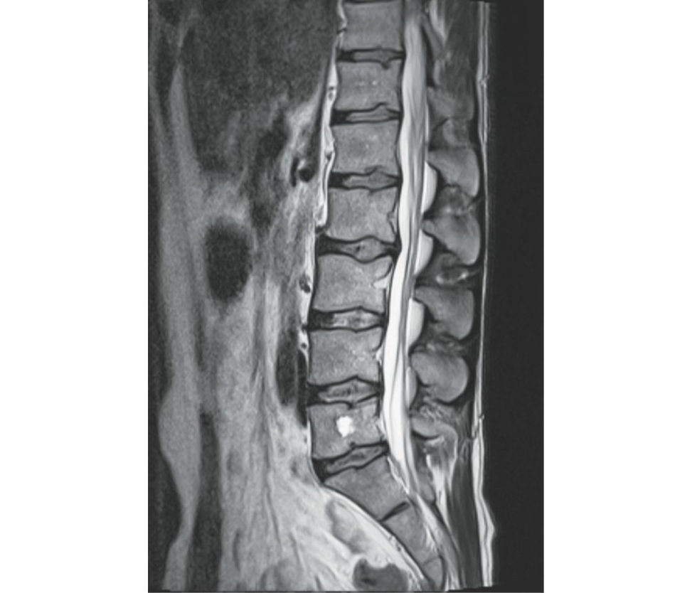
Figure 6.3a MRI lumbar spine T2 sagift al image.
T2 sagift al image (Figure 6.3a) showing hyperintense signal in the L5 body. I want to see T1, axial and parasagift al images, please.
Please comment axials at L5.
Axial T2 and T1 and fat-suppressed images (Figures 6.3b–6.3d) showing cystic mass with fluid collections in righ t-side lamina, pedicle and extending into the body of L5.
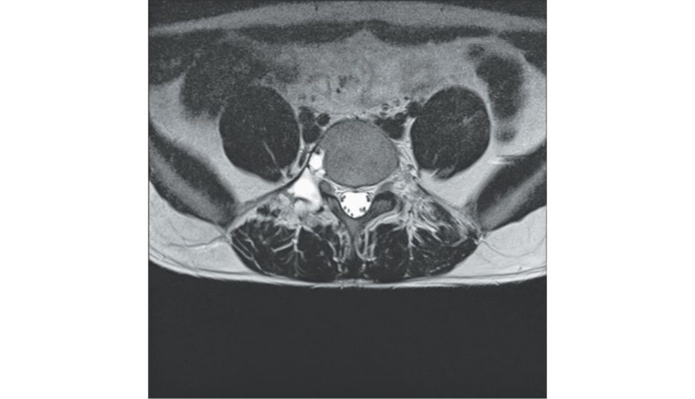
Figure 6.3b MRI lumbar spine, T2 axial image.
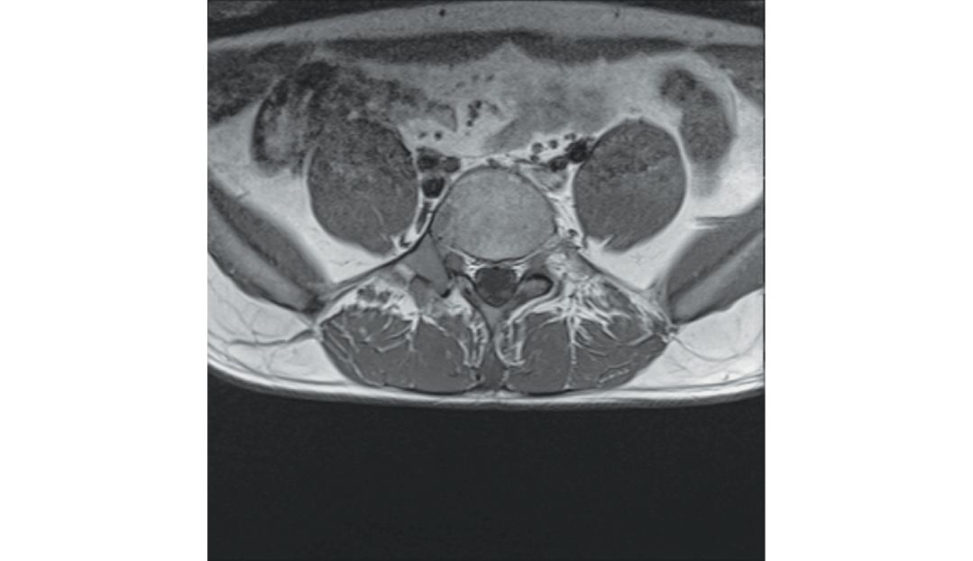
Figure 6.3c MRI lumbar spine, T1 axial image.
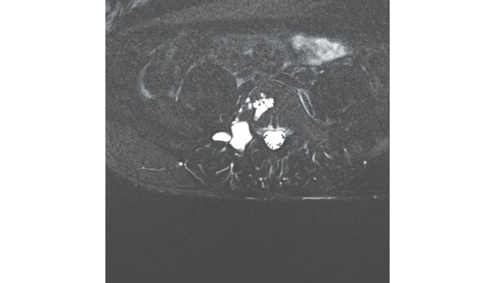
Figure 6.3d MRI lumbar spine, fat suppression axial image.
What are your differentials?
However, can’t rule this out until a biopsy is done along with culture.
What next?
I will like to refer this patient to regional oncology and the spinal team for their opinion. Also, I will get this MRI reported by a musculoskeletal radiologist.
You are the regional team.
I will get a CT scan.
Why?
Infection isles slik el yas bloods are normal and there is no fever. So, CT chest/abdomen/pelvis and bone scan for additional lesions. I need to bewary of renal cell carcinoma that can have vascular metastases that can bleed torrentially during biopsy. CT spine will also help me differentiate between ABC and telangiectatic osteosarcoma.
CT confirms this is an isolated lesion. Please comment (Figure 6.3e).
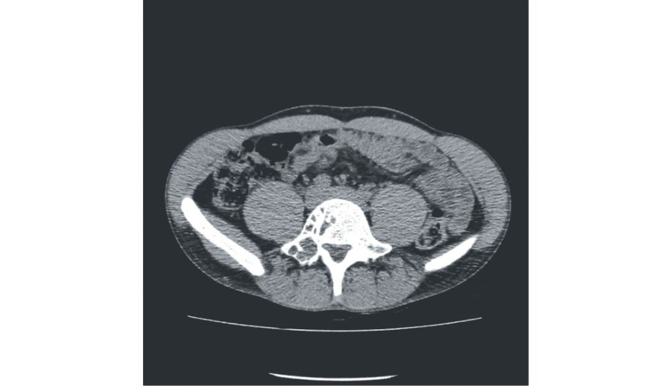
Figure 6.3e Axial CT of lumbar vertebra.
This needs biopsy to confirm it’s an ABC and not telangiectatic osteosarcoma, preferably a CT-guided biopsy.
Biopsy confirms your diagnosis of ABC. What next?
As this may lead to iatrogenic instability after resection consent for instrumentation ± fusion. Also, the patient should be counselled for recurrence as 20% of cases can recur.
Thankyou.
Learning points
1. Final confirmation of benign, malignant or infection is only by biopsy (send specimen for microbiology as well).
2. Always mention local and systemic staging for tumours.
Local – X-rays, CT.
MRI (better for soft tissue vascularity, micro metastases, intramedullary extension).
Systemic – bone scan, PET, CT chest/abdomen/pelvis.
3. Biopsy all infections and culture all tumours.
4. Know principles of biopsy.
Structured oral examination question 4#
Cervical spondylotic myelopathy
A 64-year-old man arrives in your clinic with neck pain. How would you approach this patient?
I will take a history first. Onset, duration and progress of the neck pain. Any radiculopathy?
Axial neck pain that is not severe and since the past 6 months. No radiculopathy.
Does he have any gait disturbances? Any bowel or bladder disturbances? Does he have any weakness in any of his limbs? Any red flags?
He does give a history of repeated falls. Mild clumsiness in his hands, but no bowel or bladder disturbances. No other red flags or any other significant history.
I am concerned about his repeated falls. I am thinking of cervical spondylotic myelopathy.
What next?
Proceed with examination. St art with gait and do a Romberg’s test.
He has a wide-based gait. How do you perform Romberg’s and what is its significance?
Ask the patient to stand with feet close and check whether he sways with eyes open and closed. However, if proprioception is defective, two of the sensory inputs will be absent and the patient will sway and lose balance when his eyes are closed. If he stays with eyes open, then there is a problem with the brain (cerebellum).
Romberg’s testis positive with eyes open.
I need to complete the rest of the examination.
Cervical movements are good range. Grade 3 power in hand muscles but rest all grade 5. Sensations are grossly normal except proprioception.
Reflexes in all four limbs? Hoffmans’ and Babinski’s? Any clonus?
Reflexes brisk in all four limbs. Hoffmans’ and Babinski’s are positive There is sustained clonus in the lower limbs. Rest all joints normal with good distal pulsations.
This suggests UMN lesion in all four limbs, so a lesion in brain or cervical cordLess likely a lesion in the brain, as he does not have any symptoms of the same
Brain is normal. What next?
X-rays of cervical spine and MRI scan of his full spine.
Please comment on these X-rays (Figures 6.4a and 6.4b).
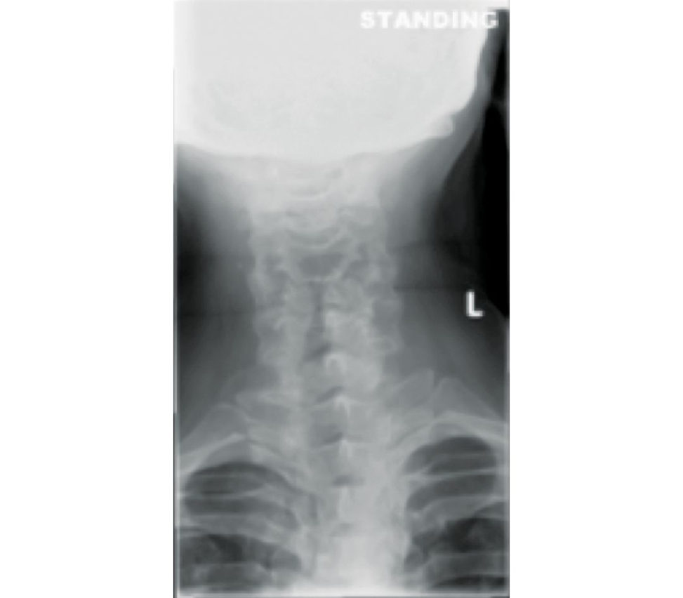
Figure 6.4a Anteroposterior (AP) radiograph lumbar spine.
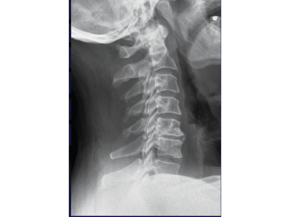
Figure 6.4b Lateral view of cervical spine.
AP and lateral view of the cervical spine showing reduced disc height, osteophyte formation at C5/6 on the lateral view. Cervical lordosis is mildly lost.
Please comment on the MRI (Figures 6.4c and 6.4d).
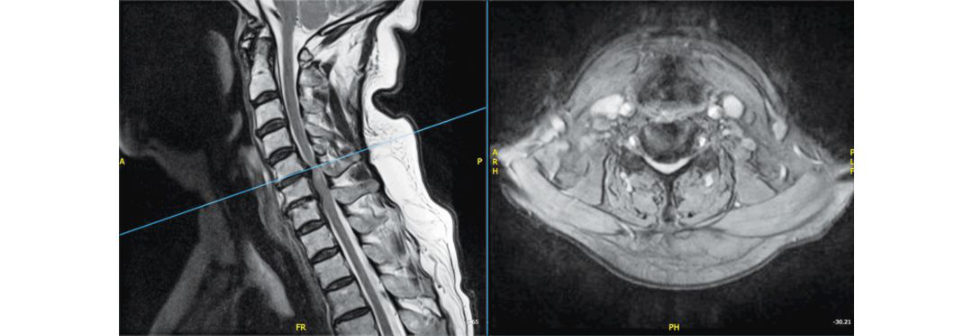
Figure 6.4c MRI (T2 sagift al and axial C5/6).
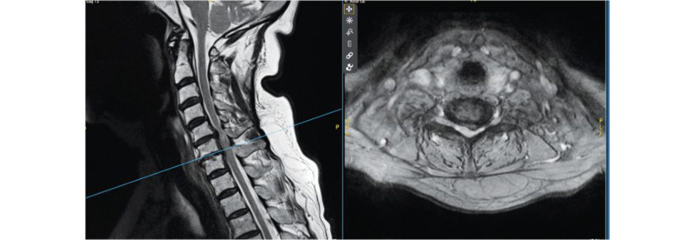
Figure 6.4d MRI (T2 sagift al and axial C6/7).
T2 MRI with axial cuts at C5/6, C6/7. There is significant compression at the disc level with cord compression. Also, I need to see T1 axial and sagift al of the cervical spine.
The rest of his spine is normal on MRI. T1 does not add much.
This is cervical spondylotic myelopathy confirmed on clinical examination and radiology. Treatment is essentially surgical as the disorder is typically progressive without surgery.
What surgery do for this condition?
As he has compression at the disc level and mainly anterior, I think he needs anterior cervical discectomy C5/6, C6/7 and fusion. This is done by the Smith Robinson approach.
What about isolated laminectomy?
Laminectomy with fusion can be done for compression mainly posteriorly or with involvement of more than three levels. But if pre-existing kyphosis is significant, then anterior or combined anterior– posterior surgery is recommended.
How would you consent this patient?
After explaining the pathology, I will tell this patient that the main aim of the surgery is to prevent the progression of the disease process.
[Interrupting] What if patient has asymptomatic c ord compression on the MRI?
close follow-up is needed with patient counselling with warning about spinal cord injury with minor trauma (e.g. central cord syndrome).
Thankyou. Learning points 1. Surgery is the mainstay for cervical spondylotic myelopathy unless the patient is unwilling or poor medically for surgery. 2. Tandem stenosis or combined cervical and lumbar stenosis can happen in 7–20% of patients Always check for upper motor neuron signs in all four limbs to pickup cervical cord compression inpatients referred for lumbar canal stenosis. 3. Smith Robinson’s approach is frequently asked for exams. 4. The role of cervical disc replacement for myelopathy. This can be performed in selected cases with compression only anteriorly, no facet arthritis, no significant neck pain. –2. 5. Causes of myelopathy. CSM (cervical spondylotic myelopathy). OPLL (ossified posterior longitudinal ligament). Infective – epidural abscess. Trauma – fractures, bilateral facet subluxation. Tumours. Congenital stenosis – Pavlov ratio < 0.8 (ratio of width of canal to width of body).
References#
1. Ikenaga M, Shikata J, Tanaka C. Long-term results over 10 years of anterior corpectomy and fusion for multilevel cervical myelopathy. Spine. 2006;31:1568–1574.
2. Gornet MF, Lanman TH, Burkus JK, et al. Cervical disc arthroplasty with the Prestige … Spine. 2017;17:1–15. doi:10.3171/2016.10.SPINE16264
Structured oral examination question 5#
Lumbar disc prolapse with caudae quina
You are on call and asked to see a 32-year-old lady with history of a fall from stairs and sustaining a fractured humerus. This fracture is closed and neurovascularly intact. There are no other injuries. How will you proceed?
I will first take a history. I will want to know mechanism of injury, any relevant past history.
[Interrupting] She says she lost balance. She has had repeated falls over the past 1 month. She is otherwise fit and well. She has been to her GP for this balance problem, who has referred her to a neurologist.
1. Does she have any hearing, visual problems? 2. Any headaches, medications cranial nerve deficits? 3. Any weakness in any of the limbs, back pain, altered sensations bowel or bladder disturbances?
She has bilateral lower limb radiculopathy. She also has bladder disturbances with reduced desire to void, straining and altered urinary sensations.
I am now worried if she has caudae quina that was probably missed in the first place. This can also explain her gait disturbances.
How does caudae quina lead to gait disturbances?
Caudae quina carries afferent inputs such as joint sense, position sense, t ouch, proprioception, etc. that provide signals to the brain to decide on balance and locomotion.
What would you like to do now?
First is the closed fracture humerus that has no neurovascular deficit and secondis the more urgent suspected caudae quina. I will like to provide analgesia, keep the patient fasting , splint the fracture and do a detailed neurological examination. I will do a per rectal examination and check for perianal sensations, anal t one. I also will request a bladder scan.
Neurology is normal in the upper limbs. Reflexes are sluggish in the lower limbs, reduced sensations equivocal plantars and motor deficit in L4–S1 dermatomes Perianal sensations are reduced, reduced anal tone. Bladder scan is not available. SLR bilaterally 30°.
Humerus fracture is undisplaced and can be managed in a plaster. It is now 4 pm and you are working in a DGH. What next?
This is caudae quina which is essentially a clinical diagnosis and needs an urgent MRI as well as lumbar spine X-rays.
Please comment on these X rays (Figures 6.5a and 6.5b).
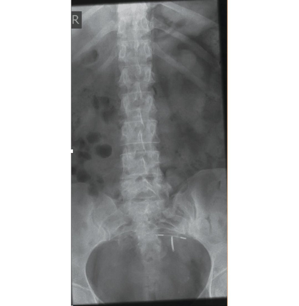
Figure 6.5a Anteroposterior (AP) radiograph lumbar spine.
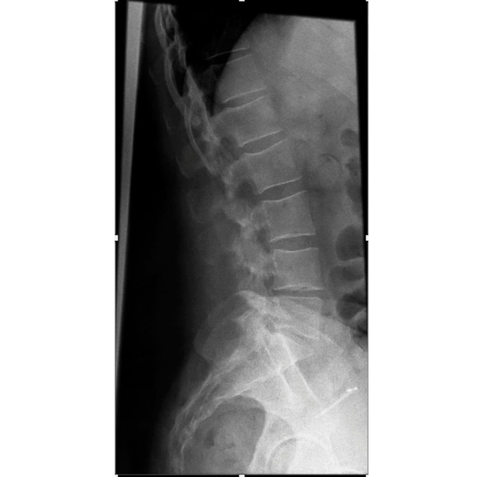
Figure 6.5b Lateral radiograph lumbar spine.
X-ray AP (Figure 6.5a) and lateral view (Figure 6.5b) of the lumbar spine. I need urgent MRI for this patient.
MRI busy but CT is available.
MR imaging is the modality of choice for caudae quina as it has greater soft -tissue sensitivity and specificity as compared to a CT scan.
Please comment on the MRI (Figures 6.5c–6.5f).
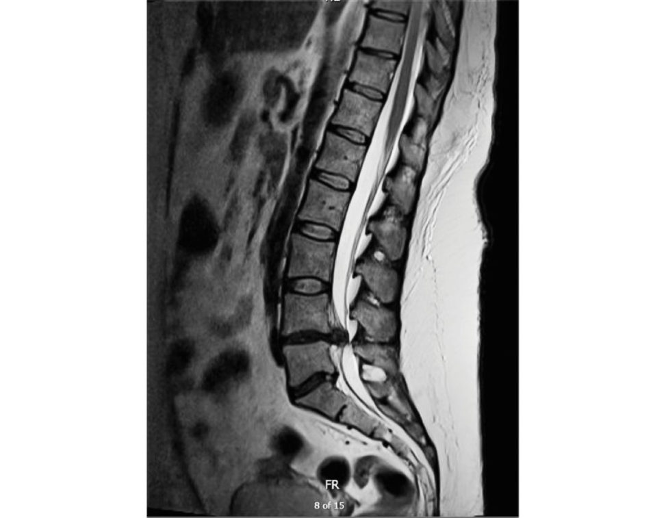
Figure 6.5c MRI sagift al T2 image.
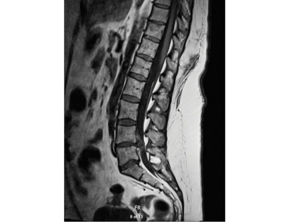
Figure 6.5d MRI sagift al T1 image.
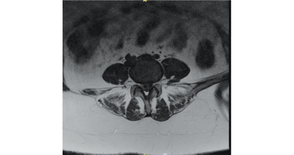
Figure 6.5e MRI axial cut lumbar spine.
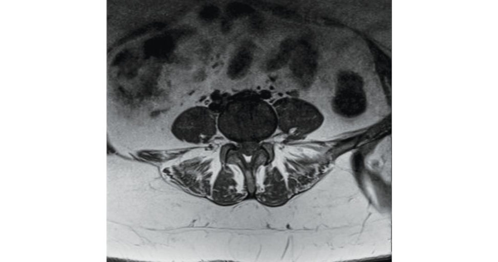
Figure 6.5f MRI axial cut lumbar spine.
MRI T2 (Figure 6.5c) and T1 (Figure 6.5d) sagift al images show a large disc at L4/5 level. The axial cuts (Figures 6.5e and 6.5f) show significant compression of the caudae quina.
What do you mean by CESI?
Management – emergency surgery. 3. CESR (caudae quina syndrome retention) – painless retention of urine with overflow incontinence. Management – emergency surgery – early CESR (< 12 hours), uncertain CESI/CESR, residual sacral nerve function present. – Next acute list – prolonged CESR. 4. CESC (caudae quina syndrome complete) – loss of all caudae quina function (absent perianal sensations patulous anus and paralyzed insensate bladder and bowel).
You mentioned emergency surgery. Would you prefer this patient to be operated on at night if it is incomplete caudae quina syndrome?
Patients operated within 24 or 48 or over 48 hafter CESI had normal bladder function in 89%, 79% and 44% of patients, respectively [2]. taking into consideration the duration of the pr e-existing symptoms and the potential for increased morbidity while operating in the small hours [3]. The principle of emergency decompressive surgery for all caudae quina is also gaining support [4,5].
Take me through the surgery for this case.
The surgical approach could be unilateral or bilateral, fenestration or laminectomy.
Anything specific regarding consent for this patient for surgery?
Besides the routine risks of discectomy, I would counsel the patient that the main aim is to prevent progression of neurology, although recovery can be expected but can’t be promised. In other words, documentation of the neurology just prior to anaesthesia would be wise from a medicolegal perspective as litigation is common in the event of persistent neurological deficit.
Thankyou. Learning points 1. Clinical assessment has low sensitivity forCES. 2. There is no agreed definition forCES, but the five characteristics are bilateral sciatica, reduced perianal sensations altered bladder function ultimately leading to painless urinary retention, loss of anal t one and sexual dysfunction. 3. Reflexes will narrow down the search for pathology into upper motor neuron lesion (brain, spinal cord) or lower motor neurons (caudae quina, peripheral nerves). So also, Babinski and Hoffmann signs are prudent to be performed.
References#
1. Todd NV, Dickson RA. Standards of care in caudae quina syndrome. Br J Neurosurg. 2016;30(5):518–522.
2. Srikandarajah N, Boissaud-Cooke MAC larkS, Wilby MJ. Does early surgical decompression in caudae quina syndrome improve bladder outcome? Spine. 2015;40:580–583. doi:10.1097/BRS.
3. Germon T, Ahuja S, Casey AT, et al. British Association of Spine Surgeons standards of care for caudae quina syndrome. Spine J. 2015;15:S2–4.
4. Bydon M, Gokaslan ZL. Time to treatment of caudae quina syndrome: a timet o re-evaluate our clinical decision. World Neurosurg. 2014;82:344–345.
5. Sonntag VKH. Why not decompress early? The caudae quina syndrome. World Neurosurg. 2014;82:70–71.
Structured oral examination question 6#
Spondylolisthesis
A 30-year-old female, otherwise fit and active presents to your clinic with a 1-year history of back pain. These are her X-rays (Figures 6.6a and 6.6b), please comment.
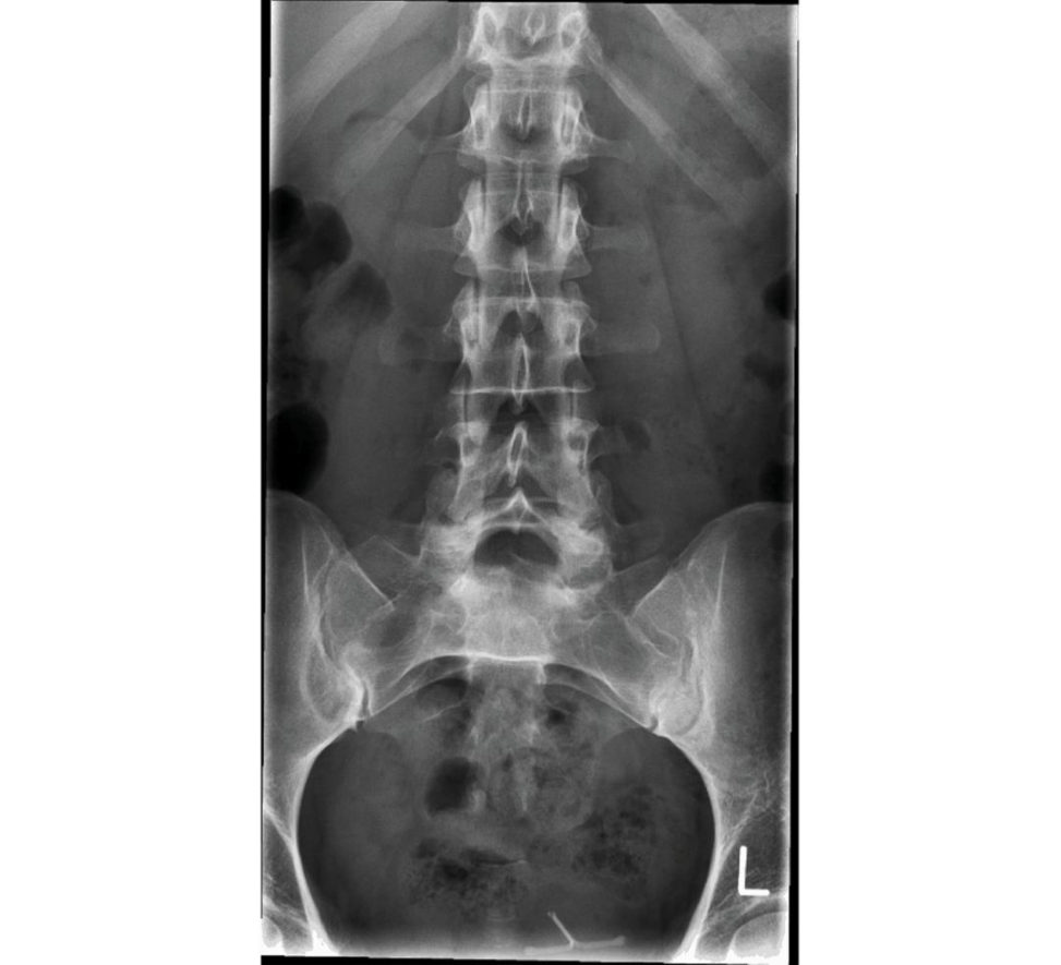
Figure 6.6a Anteroposterior (AP) radiograph lumbar spine.
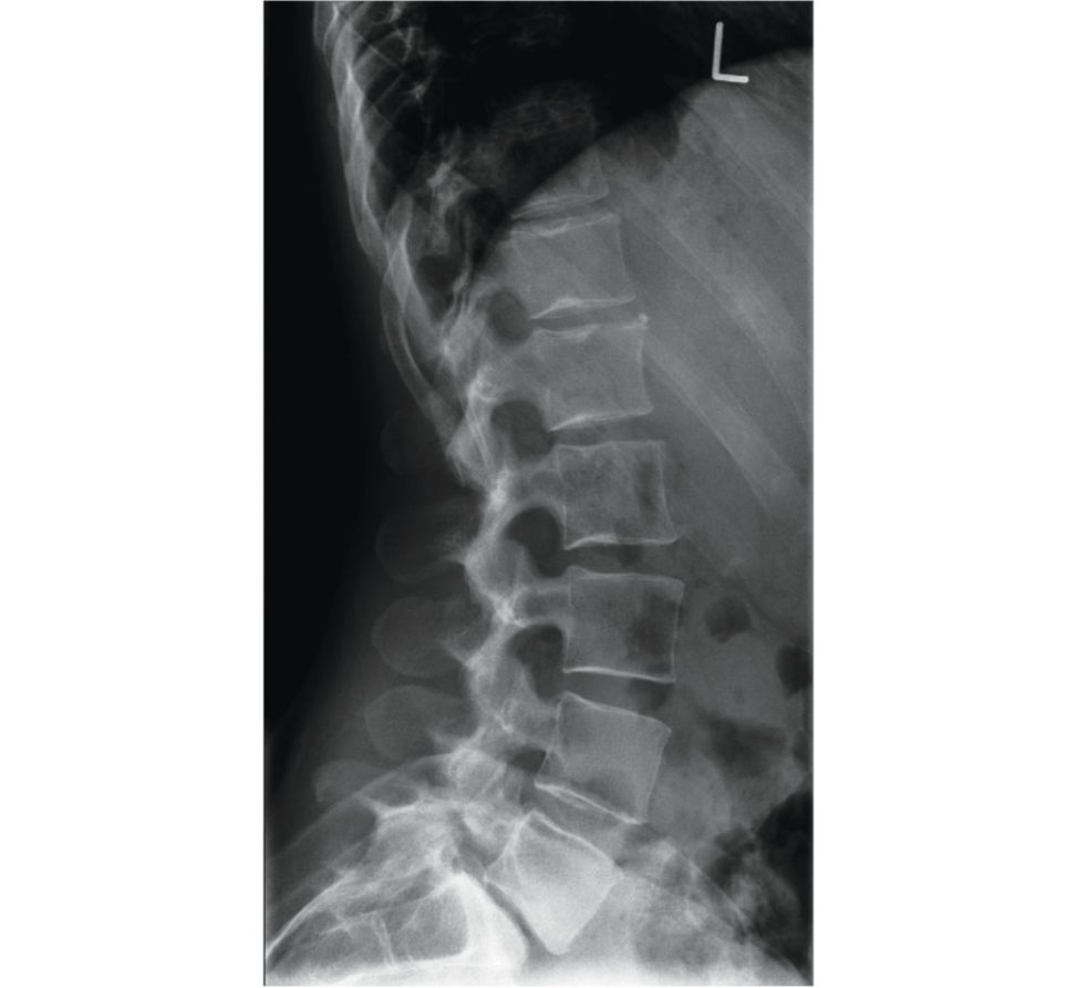
Figure 6.6b Lateral radiograph lumbar spine.
AP (Figure 6.6a) and lateral X rays (Figure 6.6b) of the lumbar spine. This is isthmic spondylolisthesis and I would say it is grade 1 according to Meyerding grading. There is also loss of disc height of the L5/S1 disc space suggesting disc degeneration. The ‘Napoleon Hat’ sign is positive (the hat is inverted with the crown representing L5 body and brim being the transverse process). Additionally , flexion–extension lateral views to look for > 4 mm translation suggesting instability.
What types of spondylolisthesis do ?
Spondylolisthesis is graded according to Meyerding’s grading system, which is graded I–V depending on the degree of displacement of the cranial vertebral body. Grade I isless than 25%. Grade II is 25–50%. Grade III is 50–75%. Grade IV is 75–100%. Grade V is spondyloptosis (> 100%).
- • IIA – pars fatigue fracture.
- • IIB – pars elongation duet o multiple healed fractures.
- • IIC – pars acute fracture.
III. Degenerative – this is due to a degenerative change that produces intersegmental instability
(due to changes in disc, joint capsules and facet joints).
IV. Traumatic – due to a fracture (but not of the pars, e.g. pedicle).
V. Pathological – caused by local bone disease (disease may not be localized).
How would you proceed?
I would like to take a detailed history, do spine examination and request further investigations His tory-wise: 1. 3. Neurology including bowel/bladder or gait involvement, any red flags. 4. Treatment she has had so far, expectations.
What radiculopathy you expect in this case? How do you differentiate symptoms from degenerative listhesis?
Degenerative listhesis is more common at L4/5 as the more sagift al-oriented facets predispose to the pathology.
This case being an isthmic spondylolisthesis at L5/S1, the patient may have either unilateral or bilateral L5 radiculopathy.
This lady has chronic back pain, L5 radiculopathy with no neurology. Her GP has exhausted all options including paint eam, activity modifications and physiotherapy. Her back pain is worse than her legs and she says she will do whatever you feel is best for her.
I need to do an examination.
[Interrupting Anything specific you want to see on examinations?
Her gait, tenderness, step sign, range of movements, nerveroot tension signs and neurology.
No neurology, ROM restricted, hamstrings are tight nothing else significant on examination. What next?
I will request an MRI scan of her lumbar spine.
Why do you need an MRI? Is the diagnosis not evident on X-rays? She also has no neurology to warrant an MRI.
This includes the anatomy and extent of compression of the L5 nerve, status of the L4/5 disc (decide on level of fusion, facet hypertrophy, ligamentum flavum hypertrophy and also whether it is a unilateral or bilateral defect).
Please comment on this MR image (Figures 6.6c and 6.6d).
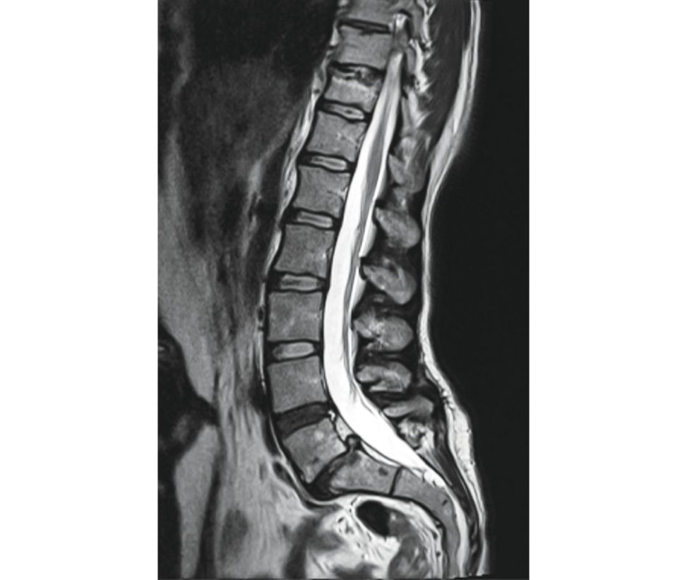
Figure 6.6c MRI T2 mid-sagift al image spine.
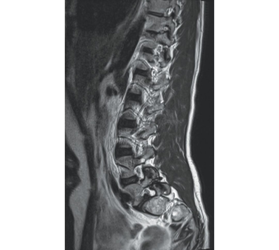
Figure 6.6d MRI para-sagift al image spine.
The T2 mid-sagift al image (Figure 6.6c) shows reduced disc height at L5/S1 with disc degeneration. The reis also disc degeneration a t the L4/5 level with a bright signal suggesting an annular tear.
Consider them not to add more information. L5 is compressed on axials. What next?
I will speak to her about surgery in the form of L5/S1 wide decompression and in situ posterolateral instrumented fusion. Factors deciding no interbody (posterolateral), TLIF/PLIF or ALIF are [1]: 1.
2. Loss of disc height.
3. Segmental lordosis.
4. Reduction of lis thesis if planned.
5. Transverse process fusion area.
disc height and lordosis is well maintained with a large area of transverse process to ensure fusion. poor transverse process fusion areas and if planning reduction.
You mentioned pelvic incidence while describing the X-rays. What do you mean by that and what is its significance?
Pelvic incidence (PI) is the angle formed between a line drawn from the centre of the S1 endplate to the centre of the femoral head and a second line drawn perpendicular to the S1 endplate intersecting it a t the centre. Low pelvic incidence indicates low shear forces at the lumbosacral junction and less lumbar lordosis. After the hips are maximally extended, further compensation is achieved with knee flexion.
In low-grade listhesis, would you recommend in situ fixation or reduction?
In situ fixation and fusion is s till the standard for low-grade listhesis. Reduction for high-grade listhesis (dysplastic isthmic) remains controversial. The risk of neurological injuries, however, can be as high as 75% [2].
Please comment on these postop X-rays (Figures 6.6e and 6.6f).
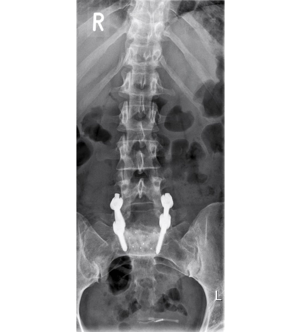
Figure 6.6e Anteroposterior (AP) lumbar spine showing in situ instrumented interbody fusion.
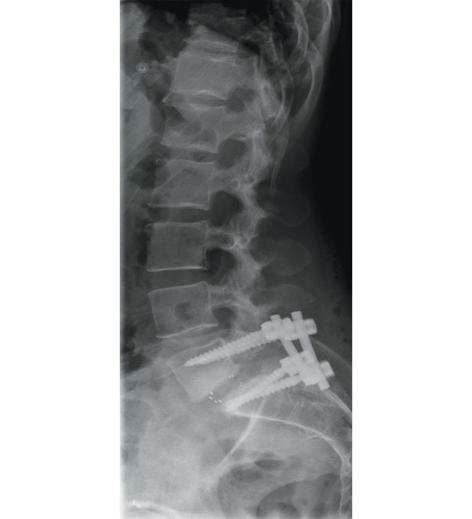
Figure 6.6f Lateral radiograph of lumbar spine showing in situ instrumented interbody fusion.
AP (Figure 6.6e) and lateral X-rays (Figure 6.6f) of the lumbar spine showing in situ instrumented interbody fusion.
This patient now has leg pain postop day one. How would you proceed?
I would again start with history, examination, investigations and management. History-wise, I would like to know if it is similar pain to that present prior to surgery. Was there any intraoperative incident such as nerve damage or dural tear?
However, if there is any neurology or significant pain, I will request an urgent CT scan first to look at screw position.
Thankyou. Learning points 1. Oblique view X-ray – discontinuity in the neck of Sc otiy dog. 2. Parsis the junction between pedicle, lamina and facet. 3. Spondylosis – defect in pars interarticularis. 4. 90% of normal volunteers will show 1–3 mm of translation on fle xion–extension lateral radiographs. More than 4 mm is regarded as abnormal. 5. Anterior retroperitoneal approach is sometimes asked for exams. 6. The above scenario can be replaced with degenerative spondylolisthesis. Know that L4/5 is the most common level. Also, it is important to know the nerveroot involved, symptoms of canal stenosis and surgical management.
References#
1. Bridwell KH. The Textbook of Spinal Surgery, 3rd edn. 2011. Lippincoft .
2. Sailhan F, Gollogly S, Roussouly P. The radiographic results and neurologic … review of 44 patientsS pine. 2006;31(2):161–169.
Structured oral examination question 7#
Ankylosing spondylitis
This is an X-ray of a 25-year-old male referred to your clinic with a history of back pain. Can you please describe this X-ray (Figures 6.7a and 6.7b)?
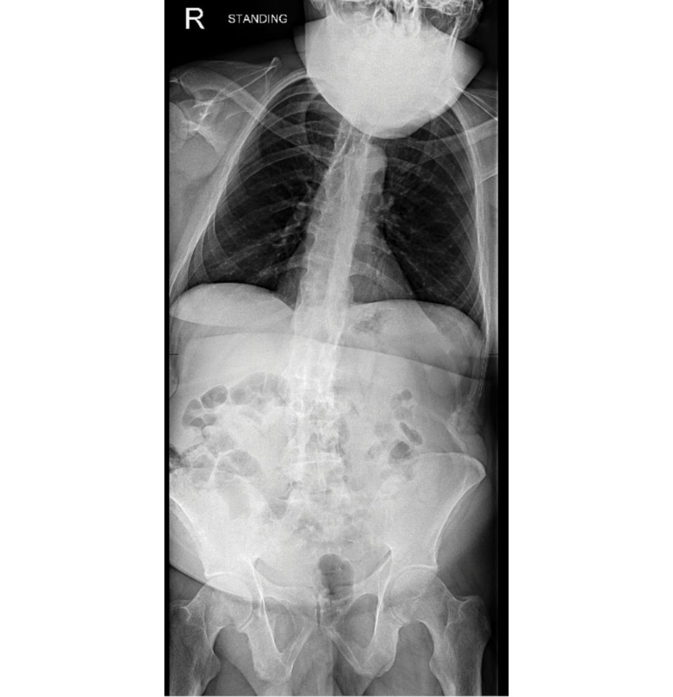
Figure 6.7a Anteroposterior (AP) full spine radiograph.
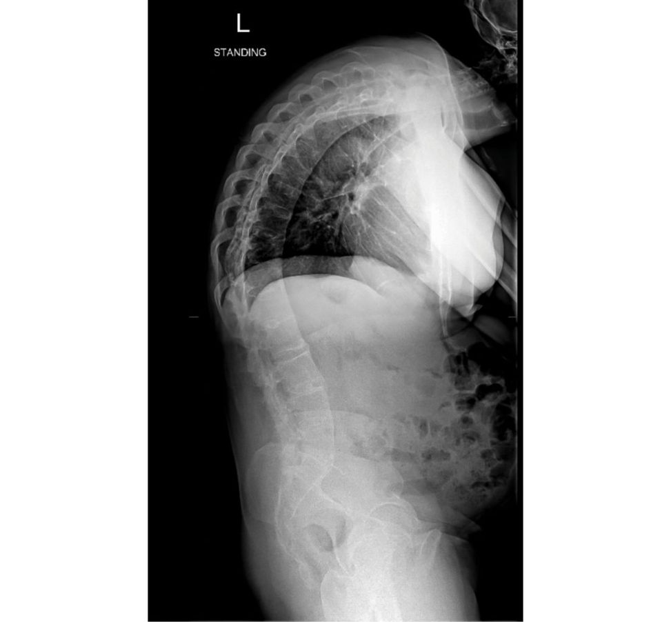
Figure 6.7b Lateral standing full-spine radiograph.
AP X-ray shows bilateral sacroiliac ankyloses and early arthritis of both hips. There is positive sagift al balance on the lateral X-rays as the C7 plumb line falls anterior to S1.
this is an X-ray of apa tien t with ankylosing spondylitis (AS)?
I need to take a history and examine this patient for obtaining my clinical criteria.
Radiological criterion
Sacroiliitis a t least grade 2 bilaterally or grade 3 or 4 unilaterally.
Clinical criteria
Low back pain and stiffness for more than 3 months that improves with exercise but is not relieved by rest.
Limitation of motion of the lumbar spine in both the sagift al and frontal planes.
Limitation of ches t expansion relative to normal values correlated forage and sex.
What are the other X-ray findings of AS?
The sacroiliac joints initially show loss of definition of subchondral bone. This is followed by marginal syndesmophytes and finally widespread ankyloses.
What is the pathology in AS?
AS is seronegative (Rh factor negative autoimmune, chronic, spondyloarthropathy. There are theories in relation to HLA B27 (90% patients are positive).
What are the orthopaedic manifestations of AS?
Bilateral sacroiliitis progressing to frank ankyloses. Other joints (knees, shoulders and ankle) – arthritis, although less likely as it mainly affects the axial skeleton.
How do you differentiate between AS and DISH (diffuse idiopathic skeletal hyperostosis)?
AS affects young males and has strong predisposition for HLA B27. Lastly, there is osteopenia in AS but increased radio density inDISH.
How would you go about examining this patient?
General examination cardiac, respiratory (reduced chest expansion, renal and abdomen). Rest of orthopaedic examination sacroiliac joint, hips, knees.
What are the hip findings in AS?
Arthritis, ankyloses, pr otusio, heterotrophic ossification and severe hip flexion deformities (usually bilateral).
If patient has concurrent spine deformity and hip deformity needing THR, what surgery would you recommend first?
As a general principle, spine deformity is addressed prior to hip. The reason suggested is that dislocation rates are higher in THR that are followed by spine deformity correction.
How do you manage these patients?
This needs a multidisciplinary approach that includes rheumatologist, physiotherapist, occupational therapist, orthopaedician. Medical management includes drugs such as NSAIDS, COX2 inhibitors and TNF alpha inhibitors.
What level are these osteotomies done?
Cervicothoracic osteotomy is performed at C7 T1 level as the vertebral artery is exterior and the canal diameter is also larger at this level.
This is a CT scan of the same patient presenting with acut e-onset back pain after a fall (Figure 6.7c). Please comment.
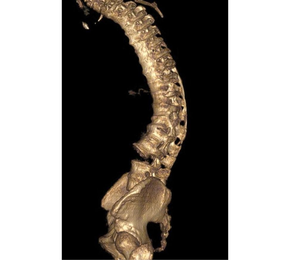
Figure 6.7c CT scan demonstrating fracture dislocation a t L3/4.
CT scan shows a fracture dislocation a t L3/4. Rest of the spine looks ankylosed.
[Interrupting] Are fractures common?
Fractures are not uncommon due to altered biomechanical properties of the spine. These fractures are invariably missed and are associated with high incidence of neurological complications and pseudoarthrosis.
Thankyou.
Other points
1. Difference between marginal (AS) and non-marginal syndesmophytes (DISH).
2. Non-articular manifestations:
Eyes: acute anterior uveitis.
Heart: conduction defects, aortis, aortic regurgitation or stenosis.
Respiratory: pulmonary fibrosis.
Renal: amyloid nephropathy.
Gastrointestinal: associated with Crohn’s disease and ulcerative colitis.
3. Preoperative anaesthesia concerns.
4. Different osteotomies for kyphosis correction.
5. Causes of kyphosis.
Structured oral examination question 8#
Idiopathic scoliosis
Could you please comment on this AP standing X-ray of a 15-year-old female who has come to your clinic with history of spinal deformity (Figure 6.8a)?
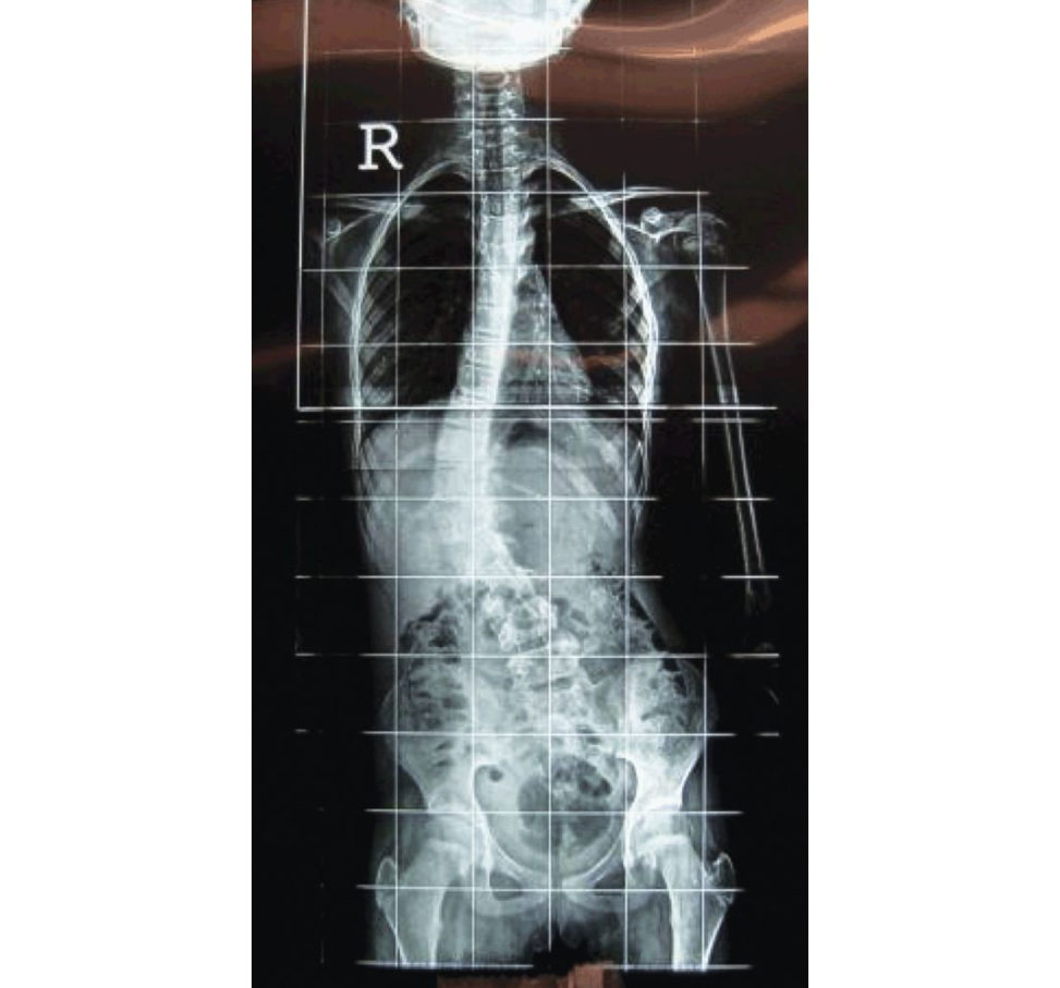
Standing PA (Figure 6.8a) spine X-ray shows a single right-sided convex thoracic scoliosis with the apex at T12. The pelvis shows Risser’s 1 stage based onless than 25% of the calcification of the lateral iliac apophysis.
What do you mean by Risser’s grading?
However, they are easily distinguished by age with Risser’s 0 at 5 years and Risser’s 5 after 16 years of age. So Risser 0 means more growth remaining and more chances of curve progression as the skeleton grows. This translates into more need of surgical intervention Rissers 5, on the other hand isless likely to progress as the skeletal maturity has been achieved.
You mentioned a Cobb angle of 50°. How is this measured?
I identified the end vertebra that has the pedicle level with the greatest tilt from the horizontal. In this case it seems to be T7 and L2.
Please comment on the lateral X-rays (Figure 6.8b).
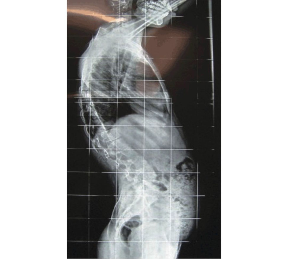
The lateral spine X-ray shows loss of thoracic kyphosis, but the sagift al balance is well maintained.
Do you feel this is an idiopathic curve?
X-ray findings suggest this is probably an idiopathic curve. But to be certain, I need to take a history, do examination and request investigations if needed.
How would you go about that?
Findings suggestive of a non-idiopathic curve are asymmetrical reflexes, neurology, foot abnormalities.
MRI features such as bone and spinal cord anomalies, syrinx, tumour or infection.
How do you differentiate between flexible and structural curve?
A flexible curve normalizes with lateral bending. The structural curve fails to correct.
What is the role of Adams’ forward bending test?
After ruling out leg length discrepancy that can be a cause of scoliosis, the patient bends forward at the waist.
Do you think this curve is likely to progress?
This is a Rissers 1 stage of skeletal growth with the curve measuring 50°. Severe curve with skeletal growth remaining is likely to progress.
Having proved this is idiopathic scoliosis on history and examination in a 12- year-old girl who has not attained her menarche, how do you proceed?
I will need to first perform lateral bending views to see if the curve corrects to any degree. This will help decide on the fusion levels.
Don’t you need an MRI?
MRI indications would be any clinical features of a generalized syndrome or foot abnormalities pointing towards a non-idiopathic scoliosis.
What next?
This is not a curve between 20 and 40° where bracing would be indicated. This curve being 50° will need surgical correction Surgical options include posterior spinal fusion, anterior spinal fusion or combined anterior and posterior spinal fusion.
Thankyou.
This is evident in the last answer with regards to management of the above patient.
Figure 6.8a Standing frontal posteroanterior (PA) radiograph spine.
Figure 6.8b Lateral radiograph spine.
Other points
1. Scoliosis is a Cobb angle measure more than 10°.
2. Define end vertebrae, stable vertebrae, neutral vertebrae and apical vertebrae.
3. Aetiology of scoliosis:
Scoliosis is a descriptive term and not a diagnosis.
(1) Idiopathic: 80% of cases.
(a) Infantile – 0 to 3 years.
(b) Juvenile – 3 to 10 years.
(c) Adolescent – 10+ years.
(2) Congenital (present at birth):
(a) Failure of formation – hemivertebrae.
b) Failure of segmentation – unilateral unsegmented bar.
(c) Mixed.
(3) Neuromuscular.
(a) Upper motor neuron: cerebral palsy.
(bLower motor neuron: polio.
(c) Muscular weakness: muscular dystrophies.
(4) Others.
(a) Syndromes: Marfans, Ehlers Danlos, neurofibromatosis.
(b) Tumours: osteoid osteoma.
(c) Trauma.
(d) Compensatory: leg length discrepancy.
4. Complications of scoliosis surgery.
5. Approaches to spine.
6. Deciding levels of fusion – include levels to correct the sagift al and coronal curvatures.
Fusion extends from the caudal end vertebrae to stable vertebrae.
Structured oral examination question 9#
Non-idiopathic scoliosis
As stated above, there are many aetiologies for non-idiopathic scoliosis.
1. Is this idiopathic or non-idiopathic? This is well answered in the previous case.
2. Is the scoliosis part of a syndrome? Look for clues such as Café-au-lait spots, axillary or inguinal freckling, Marfanoid features, scissoring gait of cerebral palsy, etc.
3. Is there any neurology? If yes, investigate with MRI (MRI is almost always needed for all non-idiopathic scoliosis) to look for cord tethering, syrinx, diatometamyelia or Arnold Chiari malformation.
4. Is the curve structural or non-structural? A non-structural curve normalizes with lateral bending.
The structural curve fails to correct less than 25°. This helps in deciding fusion levels.
5. Is the patient fit for anaesthesia? Conservative treatment with a brace for a child not fit for major surgical procedure and anaesthesia.
6. Can this scoliosis be treated with non-surgical management? Sometimes, the goal is to delay surgery to an older age. With some pathologies such as Duchenne’s muscular dystrophy, pulmonary function reduces with advancing age.
Congenital scoliosis in the form of unilateral unsegmented bar with contralateral hemivertebrae has the highest chance of curve progression (100%).
Klippel Fiel syndrome, or other systemic anomalies.
For neuromuscular scoliosis (cerebral palsy, muscular dystrophies), surgery is indicated for improvement in seating posture, trunk control or to prevent further deterioration in pulmonary function.
Because the musculature is weak, there is a good chance of recurrence if short segment fusion is done.
More so, these patients are not physiologically geared for revision surgeries or prolonged anaesthesia.
Long segment fusion from T1/T2 to sacrum (pelvic obliquity) is the usual norm for the posterior spinal fusion.
Please can you comment on these X-rays (Figures 6.9a and 6.9b)?
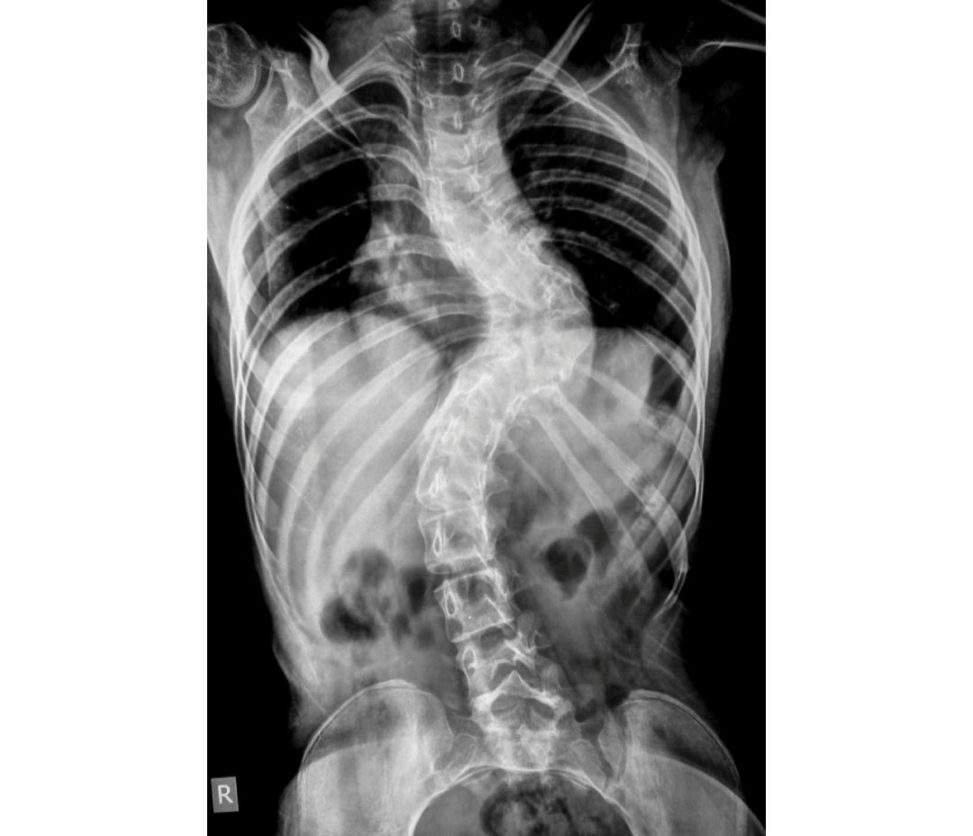
Figure 6.9a Anteroposterior (AP) radiograph of full spine.
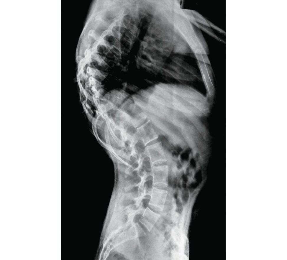
Figure 6.9b Lateral radiograph of full spine.
AP (Figure 6.9a) and lateral (Figure 6.9b) X-rays of full spine showing kyphoscoliosis of the thoracolumbar spine. The Cobb angle is about 90°.
Yes, this is neurofibromatosis (NF). So, what are the orthopaedic manifestations of neurofibromatosis?
Spine and extremities are involved in NF. Spinal involvement is the most common with scoliosis, kyphosis and atlanto-axial instability. Extremity involvement is in the form of hemihypertrophy of the limb and pseudoarthrosis of the tibia. similar to the X-rays depicted here.
How would you confirm this case to be secondary to neurofibromatosis?
According to the NIH Consensus Development Conference Statement (1987) the diagnostic criteria for NF-1 are met in an individual if two or more of the following are found: Six or more café-au-lait macules over 5 mm in greatest diameter in prepubertal individuals and over 15 mm in postpubertal individuals.
Consider this to be a case of NF in a 16-year-old female with menarche 3 years ago. The spine is progressive deformity and is the only orthopaedic manifestation. What next?
I need to take a history to know the symptoms and examine the child. I will also need further investigations.
If there is neurology, what is the cause?
The deformity itself can lead to neurology due to stretching of the neurological structures. So also, neural compromise can be due to tumour (neurofibromata).
As mentioned, this is a progressive deformity with spine the only concern. There is no neurology on examination.
I need further investigations. I need lateral bending X-rays and MRI full spine.
MRI does not add much. These are the lateral bending X-rays (Figures 6.9c and 6.9d).
There is not much correction of the curve on bending views. This being a dystrophic curve will need combined anterior and posterior spinal fusion.
Why do you need combined anterior and posterior fusion?
Pseudoarthrosis is very high with PSF alone up to the rate of 40%. Even with the combined approach, the rate is 10%.
Why not bracing?
Also, the magnitude of the curve about 90° is not suitable for bracing.
You mentioned about sagift al balance. How do you calculate that?
Sagift al balance can be found from clinical examination when viewed from the sides, but is best calculated onstanding lateral X-rays. A normal or neutral sagift al balance means a plumb line falling from the centre of C7 should fall on the posterosuperior edge of S1 vertebra. If the C7 line falls in front of S1 then it is positive sagift al balance and if it falls behind S1 then it is negative sagift al balance.
[Running out of questions] Tell me the blood supply of the spinal cord?
Blood supply is provided by: Anterior spinal artery – a single artery supplying the anterior two-thirds of the cord that includes both the lateral and ventral corticospinal tract. These inturn are reinforced by the segmental (radicular) arteries such as the ascending cervical artery, deep cervical artery, posterior intercostal arteries, lumbar arteries and lateral sacral arteries. Most often it originates from the left side between T8 and L1 vertebral segments. Damage to this artery can lead to paralysis from spinal cord infarction.
Thankyou.
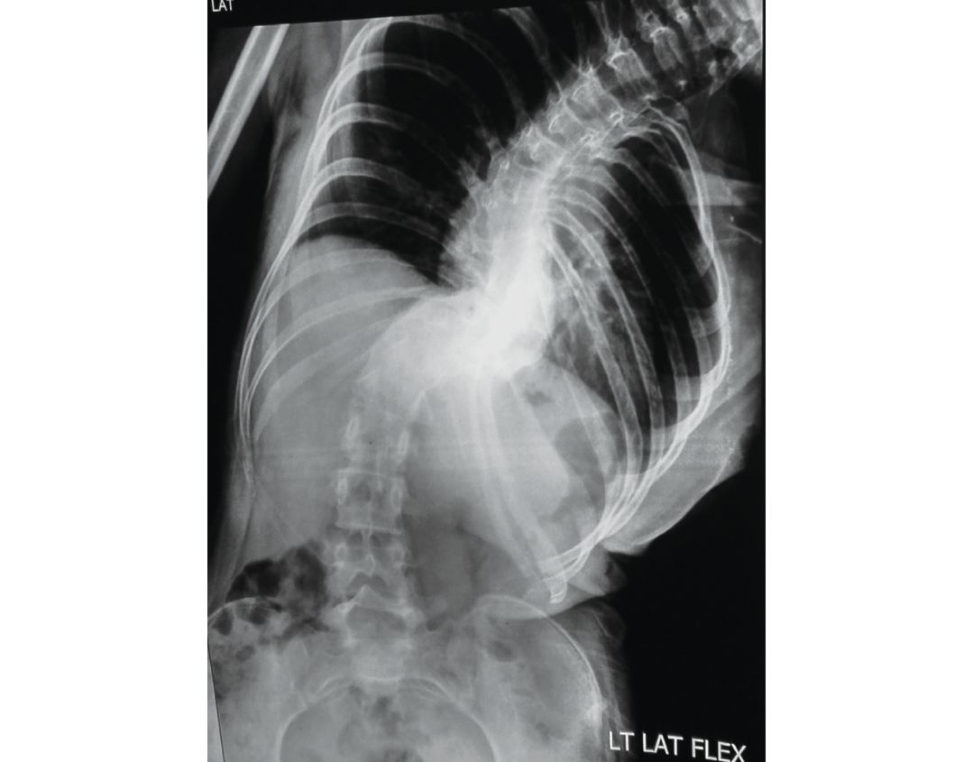
Figure 6.9c Lateral oblique bending radiographic image of lumbar spine (left lateral flexion).
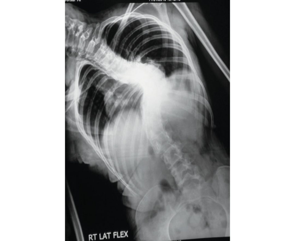
Figure 6.9d Lateral oblique bending radiographic image of lumbar spine (right lateral flexion).
Structured oral examination question 10#
Cervical prolapse disc
What changes happen in the disc with age?
Increasing age is associated with progressive dehydration of the intervertebral disc. There is an increase in the keratin to chondroitin sulphate ratio.
Can you tell me the blood supply of the adult intervertebral disc?
Adult intervertebral discs are avascular. Childre non the other hand, have vascular discs, so discitis can occur without vertebral osteomyelitis.
These are X-rays of a 35-year-old fit and healthy man working at a desk job. Can you please comment on the same (Figures 6.10a and 6.10b)?
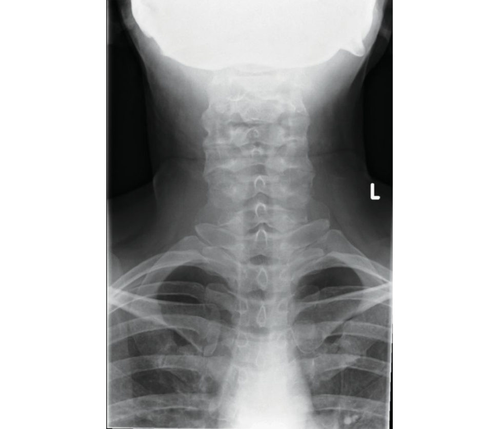
Figure 6.10a Anteroposterior (AP) radiograph of cervical spine.
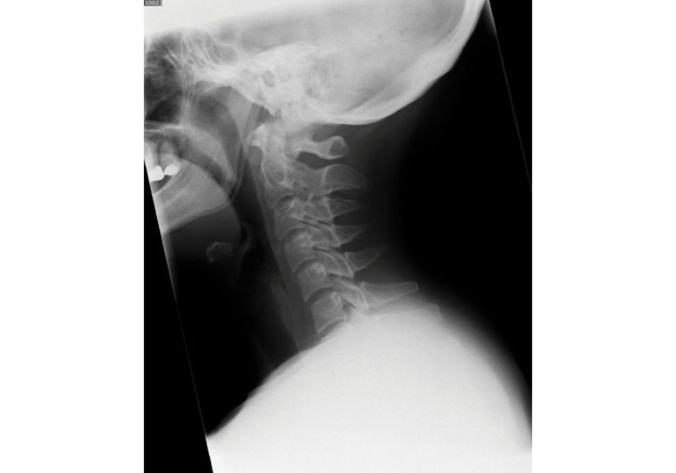
Figure 6.10b Lateral radiograph of lumbar spine.
The lateral X-ray shows loss of cervical lordosis. This lateral X-ray does not show lower cervical levels and is inadequate. The AP X-ray is largely unremarkable.
This man has right upper limb pain with axial neck pain. How would you manage this patient?
I will start with history. I would want to know how long he has had these symptoms.
His pain is along the right C7 dermatome. He is on over-the-counter analgesics. No other significant history.
I will proceed towards examination. General examination, spine examination with neurology, examination of neighbouring joints, especially the shoulders.
He has reduced neck range of motion. No neurology and no other abnormalities on examination Reflexes are normal.
Spurlings sign?
How do you perform it?
Simultaneous extension, rotation to the affected side, lateral bend, and vertical compression will reproduce symptoms in the ipsilateral arm.
This sign is positive on one side. What do you do next?
So, this patient has radiculopathy most probably due to cervical disc prolapse. I will reassure this patient provide analgesia and arrange for a follow-up in 4 weeks’ time.
Don’t you need an MRI?
His symptoms are just 4 weeks duration with no neurology or red flags. There is no indication for an MRI at this stage.
He comes back to you after 4 weeks with persistent symptoms. Now what?
I will now request an MRI.
Please comment (Figures 6.10c and 6.10d).
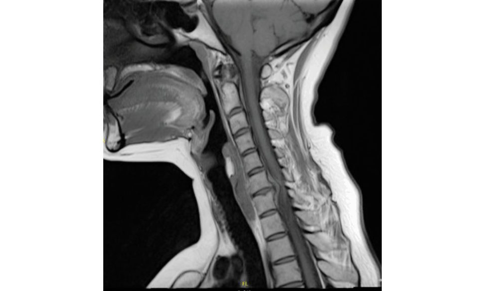
Figure 6.10c MRI cervical spine, sagift al T1 view.
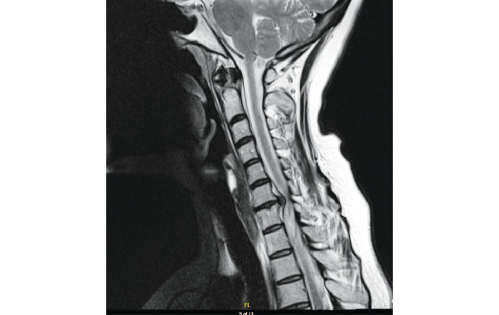
Figure 6.10d MRI cervical spine, sagift al T2 view.
T1 (Figure 6.10c), T2 (Figure 6.10d) and axial (Figure 6.10e) MR Iof the cervical spine showing a disc prolapse at C6/7 with compression of the right C7 nerveroot.
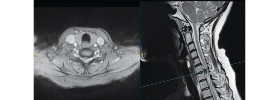
Figure 6.10e MRI cervical spine, axial views.
This patient only has symptoms of radiculopathy with no cord compression. This patient has no change of findings and is now asking questions about surgery.
I will continue with non-oper at ive management. This is as per NICE guidance for management of cervical radiculopathy 2015 [1].
Also, this is a soft disc with high water content and likely to resolve.
Lees and Turner in a long-term follow-up of patients with radiculopathy showed that 45% of patients had a single episode of radiculopathy that resolved; 30% had mild symptoms and 25% had persistent or worsening symptoms [2].
What are the surgeries for prolapsed cervical disc?
Anterior cervical discectomy and fusion, posterior foraminotomy and anterior cervical discectomy with disc replacement.
Is there any role of cervical traction?
Graham et al. found no statistically significant difference between continuous traction and placebo traction in reducing pain or improving function [ 4].
What are the advantages and disadvantages of posterior laminoforaminotomy?
It has minimal patient morbidity and avoids the need of fusion. The disadvantage is that it requires retraction of the nerveroot, risking further nerve irritation or injury.
Is it safe to offer cervical disc replacement (CDR) as compared to fusion if planning anterior surgery?
The evidence raises no particular safety issues that are not already known in relation to fusion procedures [5]. This patient is an ideal candidate for CDR if contemplating anterior surgery as the compression is mainly anterior with no posterior facet arthrosis as evident on the MRI scan.
Thankyou.
References
1. htip s://cks.nice.org.uk/neck-pain-cervical-radiculopathy
2. Lees F, Turner JW. Natural history of cervical spondylosis. BMJ. 1963;2:1607–1610.
3. Sampath P, Bendebba M, Davis JD, et al. Outcome inpatients with cervical radiculopathy: prospective multic enter study with independent clinical review. Spine. 1999; 24:591–597.
4. Graham N, Gross A, Goldsmith CH, et al. Mechanical traction for neck pain with or without radiculopathy. Cochrane Database Syst Rev. 2008;3:CD006408.
5. www.nice.org.uk/Guidance/ipg341